

Educational content from recorded physician discussions — not medical advice. Talk to your (or your child's) care team about your situation.
Video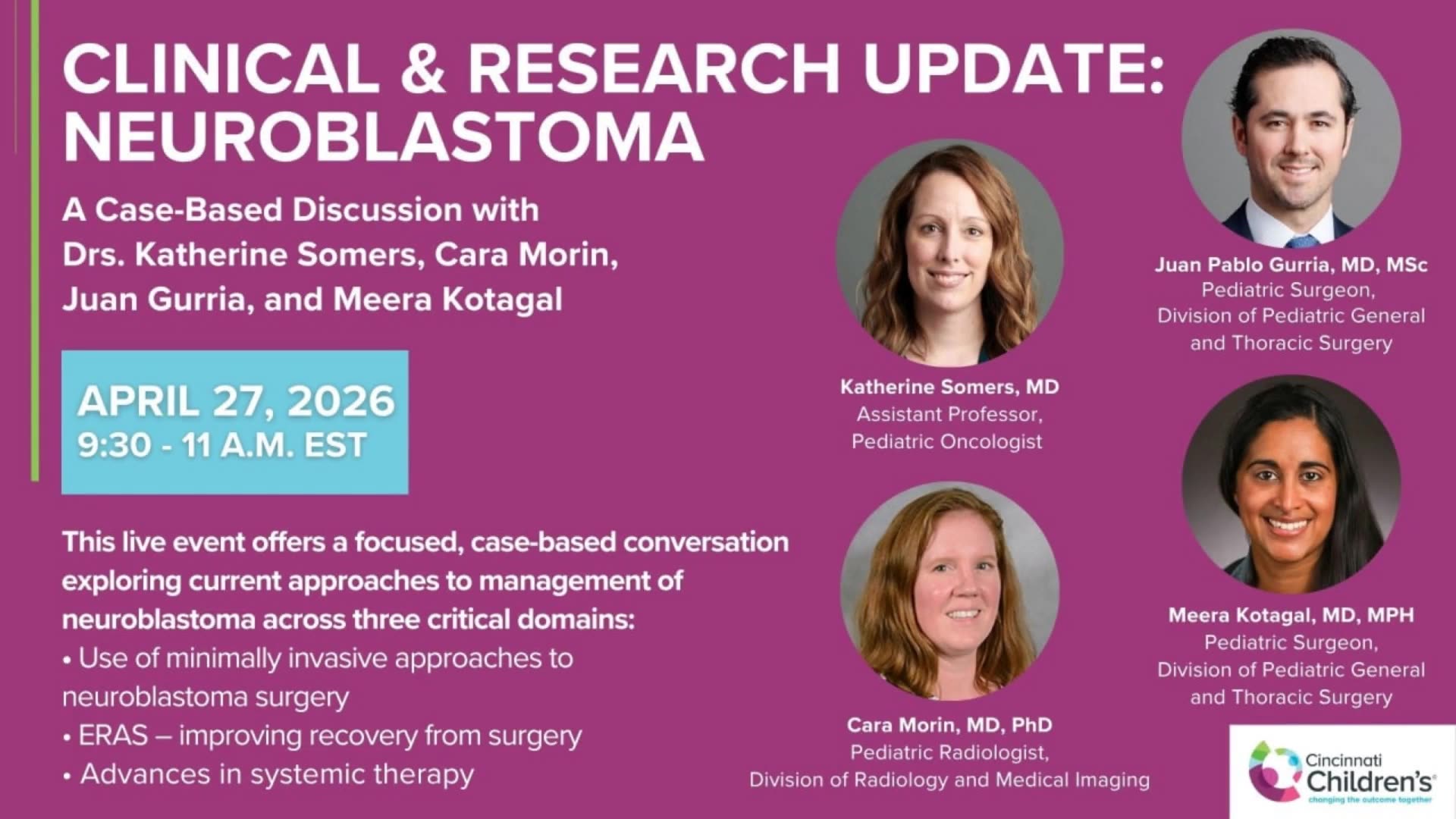
Clinical & Research Update: Neuroblastoma with Drs. Katherine Somers, Cara Morin, Juan Gurria, and Meera Kotagal
67 min · Published Sep 2026
Video
Management of Asymptomatic Lung Lesions: Pediatric Thoracic Surgery Part...
Dr. Todd Ponsky · 20 min · Published Jul 2017
Video
Postnatal Management of Lung Lesions Part III: Pediatric Thoracic Surgery...
Dr. Todd Ponsky · 27 min · Published Aug 2017
Video
Update Course 2021: TOP PUBLICATIONS IN NON - PED SURG JOURNALS
22 min · Published May 2022
Video
Image-guided core-needle or surgical biopsy for neuroblastoma diagnosis in children
54 s · Published Feb 2024
Video
Dr. Sabine Irtan - Best of the Best in Pediatric Surgery 2025
Dr. Todd Ponsky · 6 min · Published Mar 2025
Video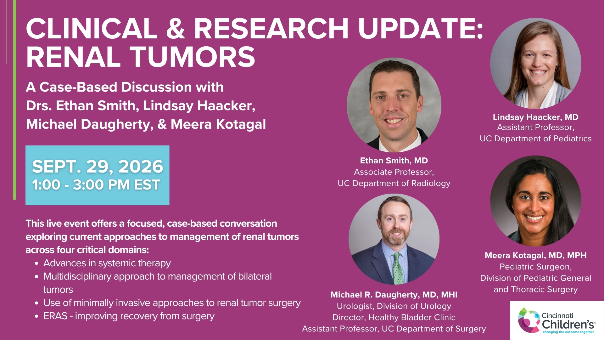
Clinical & Research Update: Renal Tumors with Drs. Ethan Smith, Lindsay Haacker, Michael Daugherty, and Meera Kotagal
71 min · Published Sep 2026
Video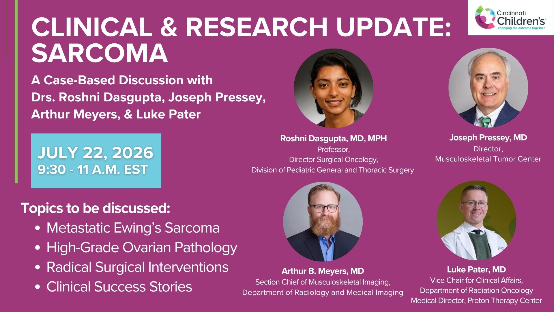
Clinical & Research Update: Sarcoma w/ Drs. Roshni Dasgupta, Joseph Pressey, Arthur Meyer, Luke Pater
66 min · Published Sep 2026
Video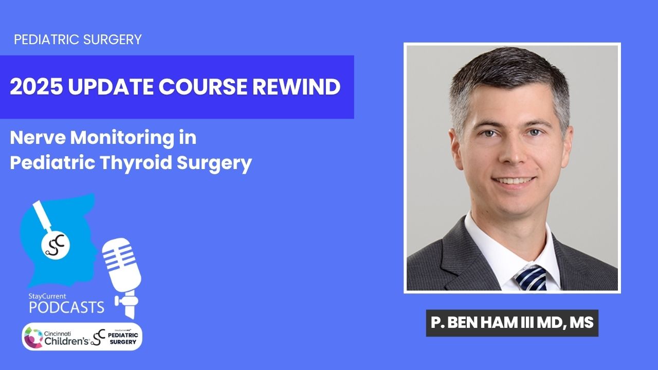
Update Course Rewind 2025: Nerve Monitoring in Pediatric Thyroid Surgery
6 min · Published Jul 2026
Video
Journal of Pediatric Surgery Article Review: June 2023, AAP Issue
Todd Ponsky · 13 min · Published Jul 2026
Video
Pediatric Surgical Oncology Research Collaborative (PSORC): Studying Rare Pediatric Tumors
56 s · Published May 2026
Video
Pooling Patients to Study Rare Pediatric Tumors: An Introduction to PSORC
56 s · Published May 2026
Podcast
Treatment for Long Gap Esophageal Atresia: Esophageal Elongation and Replacement
10 min · Published Jan 2022
Video
7th Annual Pediatric Surgery Update Course 2019 - FULL SHOW
285 min · Published Jul 2020
Podcast
Neuroblastoma
56 min · Published Aug 2019
Video
Aerodigestive & Esophageal Surgery - The Unsalvageable Esophagus & Cases
CCHMC Pediatric Surgery · 101 min · Published Nov 2018
Video
Aerodigestive & Esophageal Surgery - Difficult Tracheal Esophageal Fistula
Dr. Todd Ponsky · 120 min · Published Nov 2018
Video
Abdominal Wall Defects
37 min · Published Nov 2018
Video
Intestinal Atresia Types Explained: Grossfeld Classification for Pediatric Surgery
1 min · Published Sep 2026
Video
Derivation and validation of the Pediatric Community-Acquired Pneumonia Severity (PedCAPS) score: A prospective cohort study
50 s · Published Sep 2026
Video
FETO for Late-Diagnosed Severe Congenital Diaphragmatic Hernia (CDH) at Cincinnati Children's with Dr. Beth Rymeski
4 min · Published Sep 2026
Video
Severe Congenital Diaphragmatic Hernia (CDH) Case: FETO Management & Unexpected Findings with Dr. Beth Rymeski
3 min · Published Sep 2026
Video
Fetoscopic Endoluminal Tracheal Occlusion (FETO) Procedure: Step-by-Step Guide with Dr. Beth Rymeski
4 min · Published Sep 2026
Video
Choledochal Cyst Types Explained: Pediatric Surgery Fundamentals
1 min · Published Sep 2026
What the experts said
In younger patients, neuroblastoma is often picked up either prenatally on ultrasound, or in younger kids (two-year-old or three-year-old) as a solid abdominal mass.
When neuroblastoma patients have metastatic disease, they may present with either bony pain or potentially neurologic symptoms from cord compression.
When considering neuroblastoma as part of the differential diagnosis, it is important to get catecholamines (either urine or serum) as one of the most diagnostic laboratory tests for this tumor.
Most children with suspected neuroblastoma would get a cross-sectional imaging study, either a CT scan or an MRI.
If imaging suggests neuroblastoma (central abdominal mass or adrenal mass rather than kidney mass), the next test would be a nuclear medicine study, typically an MIBG study.
The MIBG study is helpful for confirming the diagnosis of neuroblastoma and can also demonstrate metastatic disease.
About 10% of neuroblastomas are MIBG negative.
Some centers, including Cincinnati Children's Hospital, would get a PET scan looking for tumor uptake as well as potential metastatic disease.
Based on the most recent iteration of the neuroblastoma staging system (INRGSS), it is possible to assign a stage before any invasive procedure is performed.
In the INRGSS system, tumors that are localized are categorized as L1; if localized but have image-defined risk factors (encasing nerves or vessels), they are L2; if they have metastatic disease, they are M.
There is a special category MS for children less than 18 months of age who have metastases to either the bone marrow or the skin.
The INRGSS pre-biopsy staging system was specifically created to allow studies from different centers in different countries to be compared based on the pre-surgical staging of the patient.
The prior neuroblastoma staging system required tissue diagnosis before assigning a stage.
A child with an adrenal mass on the right side and a positive MIBG scan but no evidence of metastases could potentially be treated with a primary resection of the mass via laparotomy.
Some surgeons would approach resection of a localized neuroblastoma with laparoscopy depending on the size of the tumor.
For very large masses that encase the aorta, cava, or other major vasculature, all you really want is tissue for diagnosis, which can be obtained through open biopsy, laparoscopic biopsy, or core needle biopsies done by an interventional radiologist.
The most important biologic risk determinant to obtain from neuroblastoma biopsy tissue is the NMIC status.
In addition to NMIC status, you want to look for 1P and 11Q deletions and Shimada histology in neuroblastoma biopsy tissue.
Biologic risk determinants from biopsy will tell you what risk category the patient falls into: very low risk, low risk, intermediate risk, or high risk.
Neuroblastoma risk is divided about 50-50 between the low risk categories and the high risk category, with a smaller percentage being intermediate risk.
NMIC amplification and age greater than 18 months are the most important prognostic determinants for neuroblastoma.
Patients with high risk neuroblastoma receive aggressive chemotherapy including peripheral stem cell transplant times 2, aggressive surgery with the goal of greater than 90% resection of the tumor, followed by radiation, immunotherapy after chemotherapy, and potentially retinoic acid therapy.
Intermediate risk neuroblastoma tumors get varying cycles of chemotherapy based on the biologic risk factors they have.
For intermediate risk neuroblastoma, the goal at the time of debulking or resecting the primary tumor is to achieve at least a 50% response from the initial volume of the primary tumor through the combination of neoadjuvant chemotherapy and surgical resection.
The low risk neuroblastoma group, depending on the actual age of the patient and how it is diagnosed, could potentially be followed simply with observation.
Jed Nocturne led a study through the Children's Oncology Group looking at patients less than six months of age with either a prenatally diagnosed or shortly postnatally diagnosed localized mass, showing these patients can be observed with the expectation that the vast majority will avoid any type of surgical procedure.
Patients with metastatic neuroblastoma typically receive four or five cycles of neoadjuvant chemotherapy and then are reassessed.
If the tumor and metastatic disease are responding to neoadjuvant chemotherapy, one would attack the primary tumor site with a resection, with many advocating for attempting a greater than 90% resection.
If the metastatic disease is progressing on neoadjuvant chemotherapy, then surgery is not indicated.
MS disease is specifically for patients who have a primary site with metastatic disease to the liver, the skin, or the bone marrow (specifically not bone, not cortical bone) and is less than 18 months of age.
In patients with MS disease, simple observation can be the treatment path.
If MS disease patients progress or develop respiratory issues because of an enlarging liver mass, treatment might be elected because of the complication of the size of the tumor, but the tumor itself usually does not have to be treated.
You can biopsy the skin lesions in MS disease and that will give you the diagnosis.
Neuroblastoma is the most common extracranial solid tumor in children, with the majority occurring in children less than a year old.