

From
StayCurrentMD
Image Guided Surgery Video Series, Episode 2 - Lung Nodule Localization &...
With Dr. Rajni Dasgupta · hosted by Dr. Rod Gerardo & Dr. Anton Bash & Dr. Denise Liu
Educational content from recorded physician discussions — not medical advice. Talk to your (or your child's) care team about your situation.
Video
BOB Ped Surg 2023 - Andrew Fleming, AAP - Presentation
Published Feb 2023
Video
Indocyanine green is a sensitive adjunct in the identification and surgical management of local and metastatic hepatoblastoma
1 min · Published Jan 2026
Video
Aggressive Pursuit of No Evidence of Disease Status in Hepatoblastoma Improves Survival
Published Jun 2023
Video
Social Determinants of Health Influence on Survival in Wilms Tumor, Neuroblastoma, and Hepatoblastoma
1 min · Published Jan 2026
Video
Quality of Life Outcomes for Patients Who Underwent Conventional Resection and Liver Transplantation for Locally Advanced Hepatoblastoma
1 min · Published Jan 2026
Video
Retreatment with Cisplatin May Provide a Survival Advantage for Children with Relapsed/Refractory Hepatoblastoma: An Institutional Experience
1 min · Published Jan 2026
Video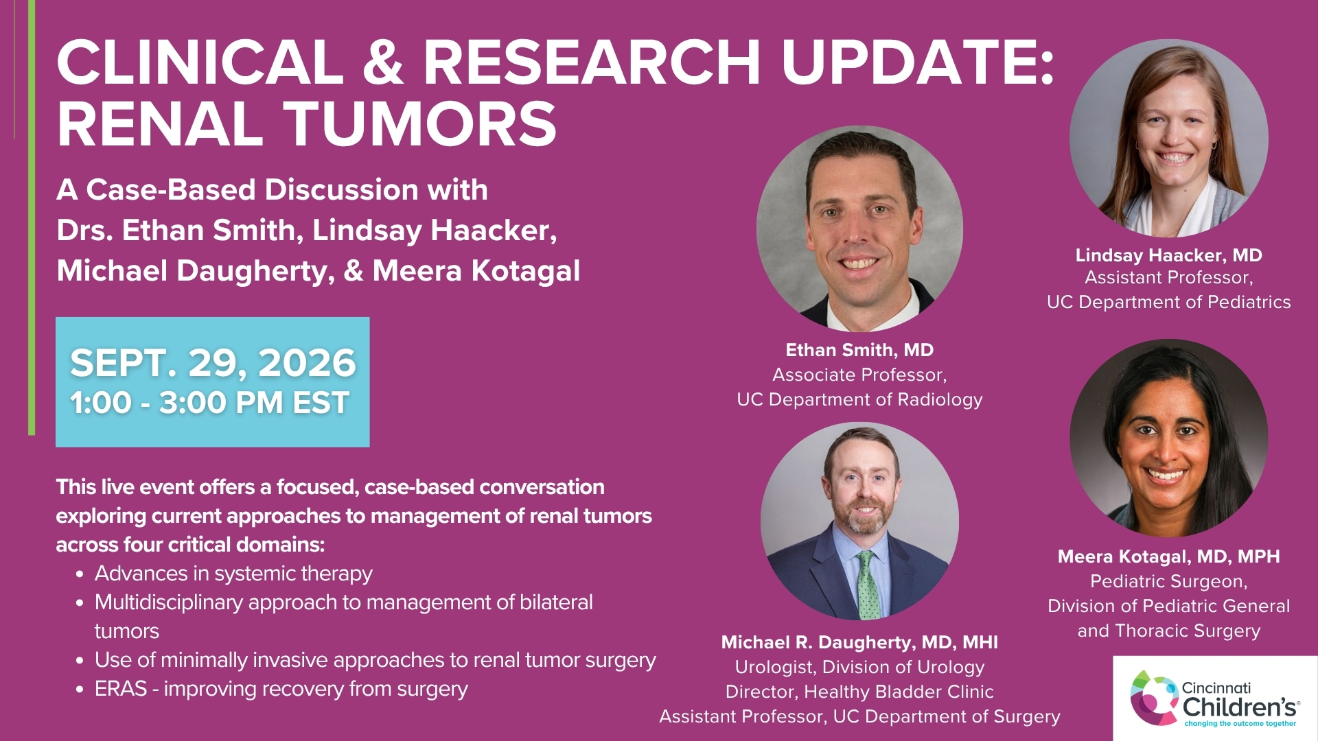
Clinical & Research Update: Renal Tumors with Drs. Ethan Smith, Lindsay Haacker, Michael Daugherty, and Meera Kotagal
71 min · Published Sep 2026
Video
Clinical & Research Update: Neuroblastoma with Drs. Katherine Somers, Cara Morin, Juan Gurria, and Meera Kotagal
67 min · Published Sep 2026
Video
Clinical & Research Update: Sarcoma w/ Drs. Roshni Dasgupta, Joseph Pressey, Arthur Meyer, Luke Pater
66 min · Published Sep 2026
Video
Update Course Rewind 2025: Nerve Monitoring in Pediatric Thyroid Surgery
6 min · Published Jul 2026
Video
Journal of Pediatric Surgery Article Review: June 2023, AAP Issue
Todd Ponsky · 13 min · Published Jul 2026
Video
Pediatric Surgical Oncology Research Collaborative (PSORC): Studying Rare Pediatric Tumors
56 s · Published May 2026
Only a few other public items share this expert — go deeper there →
Video
Intestinal Atresia Types Explained: Grossfeld Classification for Pediatric Surgery
1 min · Published Sep 2026
Video
Derivation and validation of the Pediatric Community-Acquired Pneumonia Severity (PedCAPS) score: A prospective cohort study
50 s · Published Sep 2026
Video
FETO for Late-Diagnosed Severe Congenital Diaphragmatic Hernia (CDH) at Cincinnati Children's with Dr. Beth Rymeski
4 min · Published Sep 2026
Video
Severe Congenital Diaphragmatic Hernia (CDH) Case: FETO Management & Unexpected Findings with Dr. Beth Rymeski
3 min · Published Sep 2026
Video
Fetoscopic Endoluminal Tracheal Occlusion (FETO) Procedure: Step-by-Step Guide with Dr. Beth Rymeski
4 min · Published Sep 2026
Video
Choledochal Cyst Types Explained: Pediatric Surgery Fundamentals
1 min · Published Sep 2026
What the experts said
Pre-hybrid OR, lung nodule localization was done in CT scan downstairs, then patient brought upstairs to operating room, requiring movement of the patient.
Localization typically done with Copan's breast localization needle and wire plus minus methylene blue blood patch.
More recently, localization done with microembolization coils, coils typically used for vascular embolization, advanced through Copan's needle with combination of microcoil and Copan's wire coming out in case wire becomes inadvertently pulled out during thoracoscopic resection.
Microcoil technique is very technically challenging; IR needs to practice beforehand to control the coil because it must be deployed in the right place.
Coils are smaller than guide needle for Copan; must be very careful and practice outside patient beforehand, must have holder for coils firmly established in hub, otherwise coil delivers in hub of Copan needle.
Patients always have double-lumen ET tube or bronchial blocker depending on size of patient.
Pre-hybrid OR workflow: intubate in OR, transfer patient on stretcher down to CT, get off stretcher onto CT gantry/table, do CT scan and localization, get off CT table with wire in place protecting it hoping it won't get dislodged, transfer back up to OR, position in thoracoscopic position.
There are a number of opportunities for wire to get dislodged in pre-hybrid OR workflow.
In hybrid OR, everything done more efficiently with less chance of wire becoming dislodged; when possible, position patient in thoracoscopic position to begin with so there's single prep and single draping.
Cone beam CT is a type of tomography where C-arm rotates around patient completely 360 degrees while capturing multiple images from different angles, basically like a CT but done with C-arm.
Problems have been based on anesthesia staff not understanding the procedure.
One of most important aspects of chest procedures is respiration motion; anesthesia plays crucial role in controlling respiration and giving apnea at precise moment for targeting lung lesion.
Positioning important so procedure can be done in same position as thoracoscopy without having to re-drape.
To localize lesion, go to workstation, put green line on, click on lesion with mouse, click on skin, system draws the line; advantage is line can be adjusted to go directly through middle of intercostal space above ribs to avoid arteries and nerves below rib.
System arranges C-arm automatically in exactly the right position after planning line is drawn.
Put needle into pleura to check position and stabilize with blue towels; very important to do it quickly—don't put needle in, do scan, then wait and fiddle around or you get pneumothorax or other complications.
Really important to talk to anesthesiologist and explain need for same respiratory excursion whenever doing cone beam CT imaging, and that has to correspond to when actually advancing needle.
If cone beam CT done with full inspiration, location of nodule will be different in 3D than if done in full expiration; planned paths are planned from respiratory excursion from first CT, so unless same respiratory excursion when advancing needle, will be off target.
Even if complication occurs, most common one being pneumothorax, it's not a disaster—just put 5 French sheath or catheter and connect to low wall suction, which will take care of pneumothorax.
For lung biopsy, nodule has to be at least 1 centimeter because chasing after lung nodule is difficult.
Cone beam CT can see nodules like traditional CT; for big nodules, turn dose down and use enough X-ray dose to get job done.
Cone beam CT has lung optimization module; able to see every small lesion seen on traditional CT downstairs diagnostically beforehand in hybrid OR upstairs.
Contrast resolution and spatial resolution of cone beam CT is just as good as traditional CT, in fact slightly better.
Can localize multiple lesions at same time; up to 3 have been done.
When doing multiple localizations, increased chance of pneumothorax, but just put small catheter in and suck it out.
If lesions all within same lobe, thoracic surgeon can take it all out; if in different lobes and worried they're of different etiology, can localize them, but usually just pick biggest one.
Can give nodule segmentation coloring (e.g., little green dot) and do 3D rendering; can rotate image around in 3D space to understand nodule position relative to needle tip.
Actual imaging is super helpful, but also helpful to have interventionists in room for real-time discussion of needle position (medial, anterior, posterior) and confirmation.
Radiologists and surgeons think slightly differently in 3D space; good to have trusted colleagues in all areas discussing together so it's very clear.
Interventionalists stay in room while positioning and surgery are done, or come back to room fairly frequently if must do something else.
Goal is to prep and drape once; rare occasion may require supine position rather than lateral decubitus depending on nodule location, but that's relatively rare.
Limiting changing and movement helps ensure wire stays in place and saves time.
Ideally, as advancing through parietal pleura and puncturing visceral pleura, inject a little methylene blue blood patch going down toward target.
For very peripheral nodules, if don't have decisive puncture through visceral pleura and are injecting fluid/methylene blue blood patch, can push away visceral pleural surface of lung; think you've got deep enough but haven't actually punctured through visceral pleura, so when deploy wire, wire won't be within lung.
Must be decisive when puncturing visceral pleura to avoid wire deployment outside lung.
Radiographs are a shot in time, fluoroscopy gives real-time 2D images, and latest fluoroscopic machines can do cone beam CT which performs 360-degree scan of designated 3D volume, giving cross-section images.
Localization on CT is simple: draw line between skin surface and target on CT images; beauty of cross-sectional CT images is you can account for z-axis, giving 3D rendition or 3D space.
To ensure same respiratory effort, ask anesthesia colleagues to fully paralyze patient and disconnect tubes from ventilator so patient can exhale naturally and stop at very natural stopping point—most reproducible rather than having CPAP of 20 or 25.
Having no positive pressure reduces chance of pneumothorax.
If lesion is peripheral, high risk of pneumothorax; if lesion is more central, higher risk of pulmonary hemorrhage.
Under plain fluoroscopy, nodule that small can't be seen, so would be pushing blind without CT and planned graphics.
Display monitor shows live X-ray image with graphics and pre-op CT so can compare all images while doing procedure.
3D rendering takes time; it's a tool in the armamentarium but may not be needed for every case.
Added benefit from technologist perspective: having surgeon and interventional radiologist in room helping staff physician so everyone's happy when they get in there.