

From
StayCurrentMD
Image Guided Surgery Video Series, Episode 2 - Lung Nodule Localization &...
With Dr. Denise Liu · hosted by Dr. Em Gootee & Dr. Rod Gerardo & Dr. Anton Bash
Part of
Pediatric Oncology 692 items
Educational content from recorded physician discussions — not medical advice. Talk to your (or your child's) care team about your situation.
Only a few other public items share this expert — go deeper there →
Video
Intestinal Atresia Types Explained: Grossfeld Classification for Pediatric Surgery
1 min · Published Sep 2026
Video
Derivation and validation of the Pediatric Community-Acquired Pneumonia Severity (PedCAPS) score: A prospective cohort study
50 s · Published Sep 2026
Video
FETO for Late-Diagnosed Severe Congenital Diaphragmatic Hernia (CDH) at Cincinnati Children's with Dr. Beth Rymeski
4 min · Published Sep 2026
Video
Severe Congenital Diaphragmatic Hernia (CDH) Case: FETO Management & Unexpected Findings with Dr. Beth Rymeski
3 min · Published Sep 2026
Video
Fetoscopic Endoluminal Tracheal Occlusion (FETO) Procedure: Step-by-Step Guide with Dr. Beth Rymeski
4 min · Published Sep 2026
Video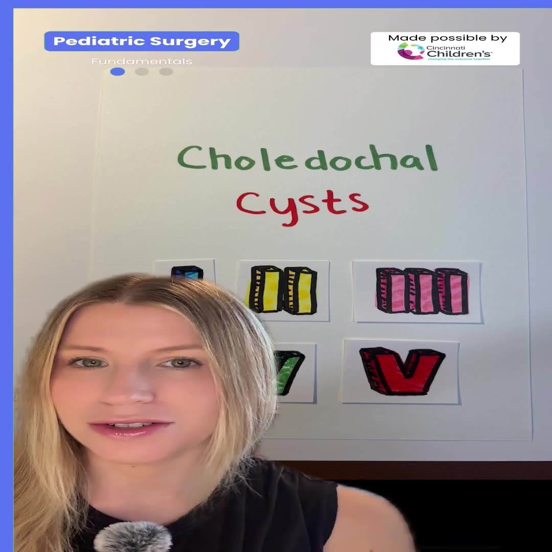
Choledochal Cyst Types Explained: Pediatric Surgery Fundamentals
1 min · Published Sep 2026
What the experts said
The localization procedure from patient entry to surgical readiness takes approximately 45 minutes total.
The surgeon retrieves the localization wire into the chest before lung deflation to prevent wire dislodgement during lung collapse, as the wire can become stuck in chest musculature.
The localization wire is cut at skin level after CT-confirmed placement because the wires are quite long and cutting facilitates surgical handling.
Cutting the wire at skin level prevents inadvertent removal during patient repositioning and prevents the wire from bending or jamming at the skin surface during transport.
In one historical case, OR nurses unfamiliar with the wire localization mistook the wire for a retained stitch and removed it, prompting the current protocol of cutting wires short.
Fluoroscopy during resection is used only when a coil has been placed, to confirm the coil has been removed with the specimen and is not retained in the patient.
Indocyanine green (ICG) is metabolized by the liver, making it useful for visualizing hepatoblastoma metastases.
Enhancement of pulmonary nodules with ICG depends on the primary cancer's vascularity; sarcomas show hyperenhancement due to increased angiogenesis.
ICG is not specific for tumor type because multiple primary cancers can be hypervascular.
ICG has limited depth of penetration, restricting visualization of deep lung lesions thoracoscopically; the specimen often must be opened to confirm nodule removal.
Needle localization is important for nodules not at the lung periphery because ICG cannot reliably visualize them thoracoscopically.
At Cincinnati Children's, interventional radiology moved from the radiology department into the operating room in 2000.
Interventional radiology procedures are scheduled on the operating room schedule with OR anesthesia, making IR part of the operating room rather than radiology.
Physical integration of interventional radiology into the operating room is uncommon; few hospitals have achieved this despite attempts.
The surgical approach uses three ports total: one camera port and two working ports.
The surgeon does not initially insufflate the chest, finding and retrieving the wire while the lung is inflated before working with anesthesiology to deflate the lung.
Wedge resection is performed using a thoracoscopic stapler, and tissue sealant is applied over staple lines to prevent bleeding.
The specimen is radiographed after removal to verify complete removal of wire or coil.