

From
Live Event Content
2025 Pediatric Surgery Update Course - Updates in Central Line Management
With Dr. Nathan Fagan
Educational content from recorded physician discussions — not medical advice. Talk to your (or your child's) care team about your situation.
More from this expert
Dr. Nathan FaganVideo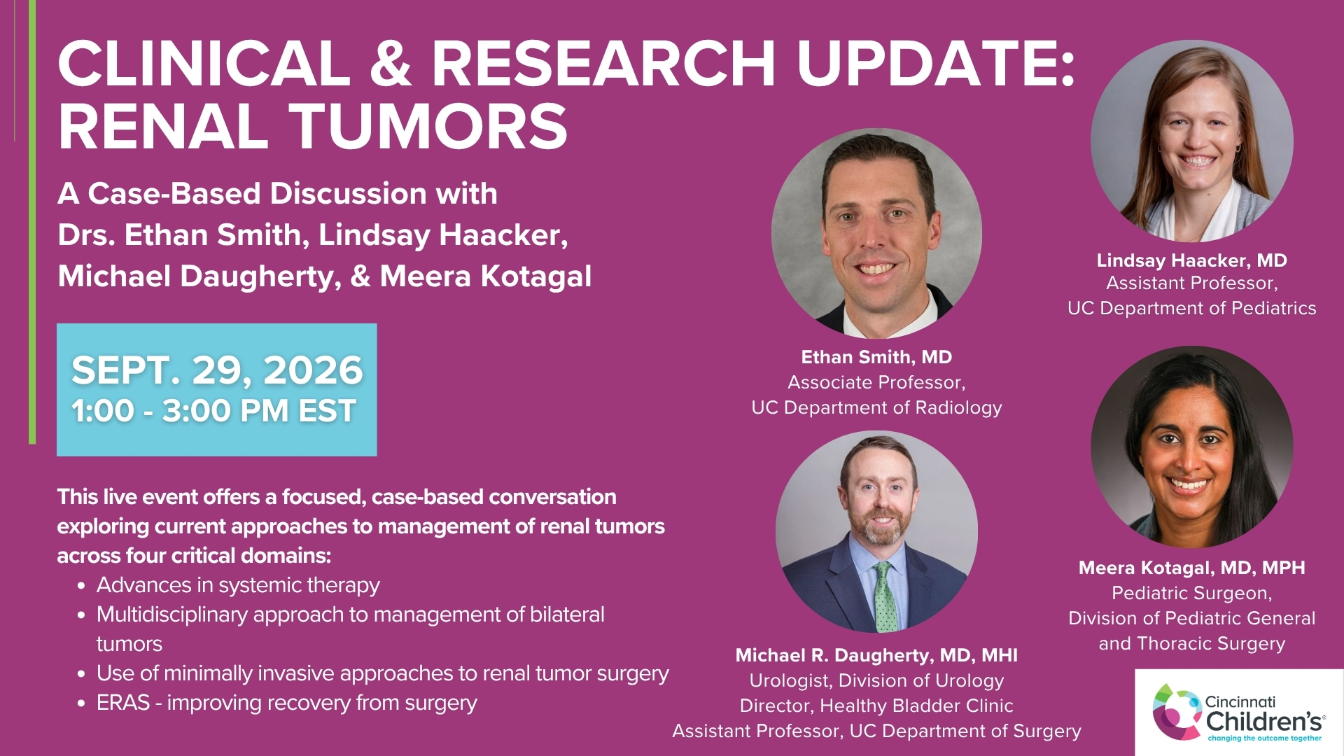
Clinical & Research Update: Renal Tumors with Drs. Ethan Smith, Lindsay Haacker, Michael Daugherty, and Meera Kotagal
71 min · Published Sep 2026
Video
Clinical & Research Update: Neuroblastoma with Drs. Katherine Somers, Cara Morin, Juan Gurria, and Meera Kotagal
67 min · Published Sep 2026
Video
Clinical & Research Update: Sarcoma w/ Drs. Roshni Dasgupta, Joseph Pressey, Arthur Meyer, Luke Pater
66 min · Published Sep 2026
Video
2026 Laparoscopic Pediatric Hernia Repair
123 min · Published Jun 2026
Video
Beyond the Spectrum: Diagnosis, Myths & Management - Caitlin Couch & Leslie Lopez - APP Conference 2026
50 min · Published May 2026
Video
Dysautonomia: Navigating the Journey - Martha Willis - APP Conference 2026
55 min · Published May 2026
What the experts said
A teenager with a left subclavian dual-lumen port had a history of subclavian thrombus successfully treated with heparin, but the medial lumen would not flush while the lateral lumen functioned normally.
The non-flushing port was found to be simply not accessed correctly; re-accessing it allowed it to flush normally without any other intervention.
Removing and replacing a port for access issues without fluoroscopic evaluation risks losing potential future access sites and may contribute to the cascade creating 'vasculopath' patients with no upper or lower central access.
Sharp recanalization of the SVC can be performed using ultrasound to visualize the jugular vein and a combination of fluoroscopy and ultrasound to guide a small needle through the stenosis into the right atrium.
Two techniques for sharp recanalization include: (1) using a contrast-filled circle as a target from above while advancing a needle from the femoral vein below, and (2) inflating a contrast-filled balloon from below as a target for needle puncture.
The thoracic azygos approach for central access involves staying anterior and lateral to the carotid artery, using ultrasound guidance, and is described as similar to laparoscopic inguinal hernia repair in technique.
The Surface EKG system by Merit Medical allows image-guided SVC recanalization by advancing a catheter from the femoral vein to the occlusion site, defining a target exit point, and driving a needle through the catheter to create through-and-through access.
The Surface EKG technique works best when there is at least a remnant of vessel present to traverse, rather than complete chronic occlusion.
Intracardiac echocardiography (ICE) catheters project ultrasound waves 360 degrees and can reconstruct images in any plane, potentially allowing safer guidance of needles across vascular occlusions.
Traditional ICE catheters are 10 French, newer versions are 8 French, and some can be used in 7.5 French sheaths; 10 French is difficult in pediatric patients but 8 French is more feasible.
When dilating across tight vascular occlusions, balloons are used rather than traditional dilators, starting with small cardiac balloons (5mm) and progressively increasing size.
Transhepatic access may be easier to control if bleeding occurs or the catheter dislodges, particularly in pediatric patients, but weak evidence suggests higher thrombosis rates compared to translumbar access.
Two patients with transhepatic catheters experienced dislodgement into the peritoneal cavity due to respiratory movement, resulting in TPA infusion into the abdomen and severe complications.
Translumbar catheter dislodgement results in retroperitoneal bleeding which may be contained by muscle, whereas transhepatic dislodgement causes hemoperitoneum.