

Educational content from recorded physician discussions — not medical advice. Talk to your (or your child's) care team about your situation.
Only a few other public items share this diagnosis — go deeper there →
Video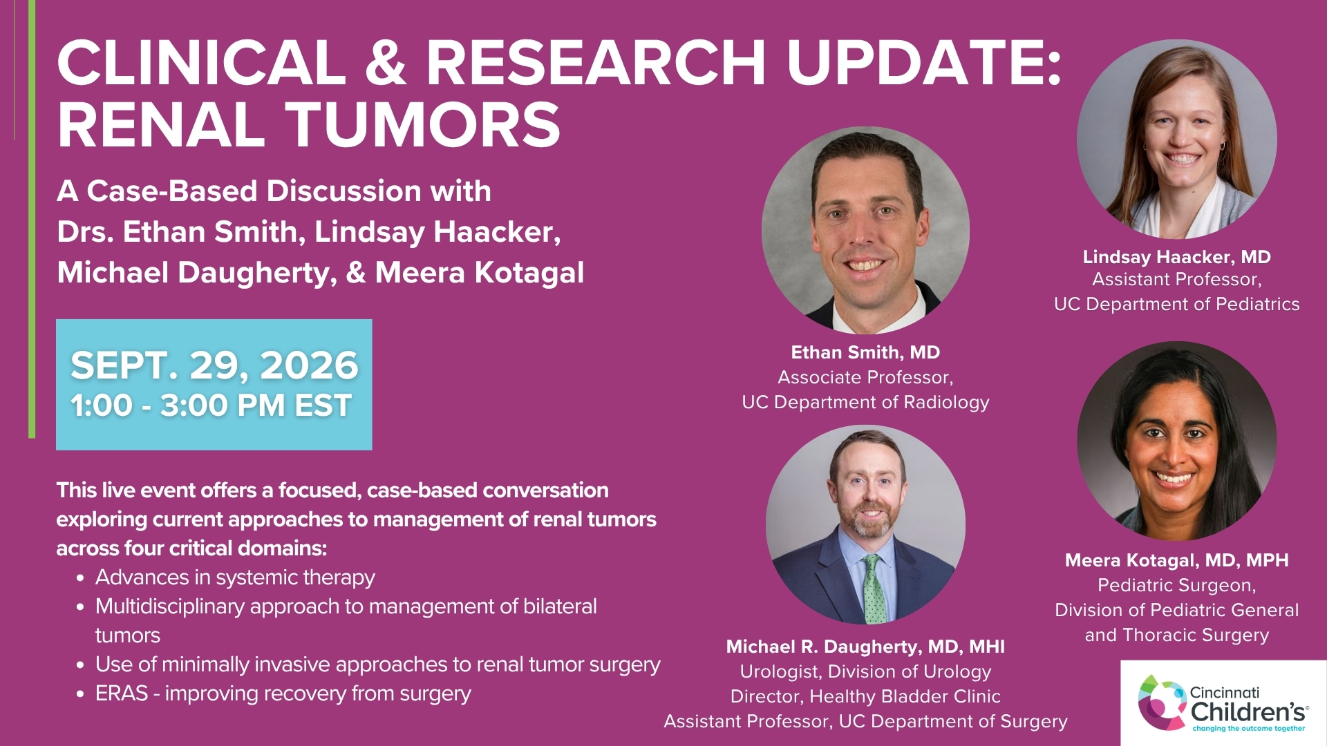
Clinical & Research Update: Renal Tumors with Drs. Ethan Smith, Lindsay Haacker, Michael Daugherty, and Meera Kotagal
71 min · Published Sep 2026
Video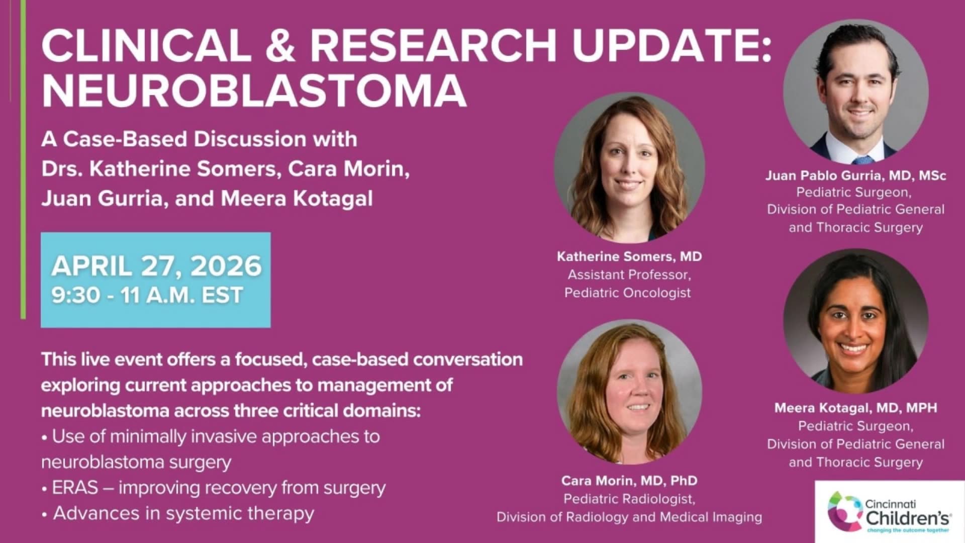
Clinical & Research Update: Neuroblastoma with Drs. Katherine Somers, Cara Morin, Juan Gurria, and Meera Kotagal
67 min · Published Sep 2026
Video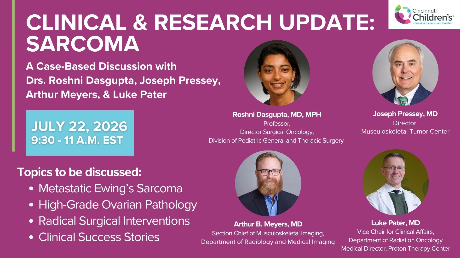
Clinical & Research Update: Sarcoma w/ Drs. Roshni Dasgupta, Joseph Pressey, Arthur Meyer, Luke Pater
66 min · Published Sep 2026
Video
Update Course Rewind 2025: Nerve Monitoring in Pediatric Thyroid Surgery
6 min · Published Jul 2026
Video
Journal of Pediatric Surgery Article Review: June 2023, AAP Issue
Todd Ponsky · 13 min · Published Jul 2026
Video
Pediatric Surgical Oncology Research Collaborative (PSORC): Studying Rare Pediatric Tumors
56 s · Published May 2026
Video
sealer demo
Dr. Steve Rothenberg · 3 min · Published May 2026
Video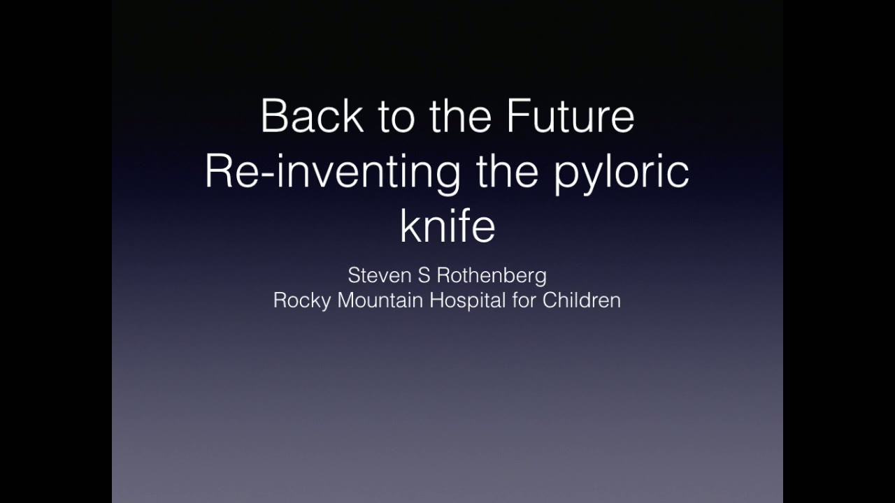
pyloric knife
Dr. Steve Rothenberg · Published May 2026
Video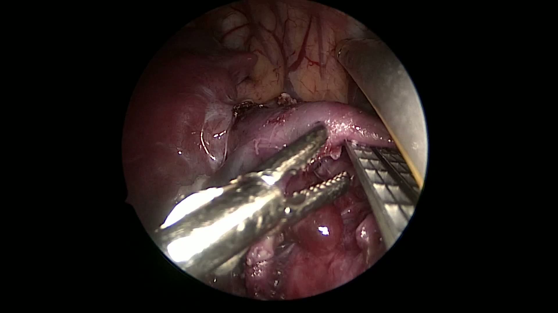
pulm artery LLL
Dr. Steve Rothenberg · 9 s · Published May 2026
Video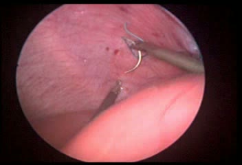
plication narrated
Dr. Steve Rothenberg · 6 min · Published May 2026
Video
percuvance redo nissen
Dr. Steve Rothenberg · 5 min · Published May 2026
Video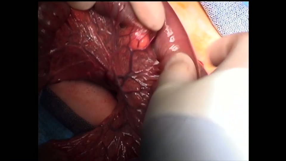
open bowel anast
Dr. Steve Rothenberg · Published May 2026
Video
Intestinal Atresia Types Explained: Grossfeld Classification for Pediatric Surgery
1 min · Published Sep 2026
Video
Derivation and validation of the Pediatric Community-Acquired Pneumonia Severity (PedCAPS) score: A prospective cohort study
50 s · Published Sep 2026
Video
FETO for Late-Diagnosed Severe Congenital Diaphragmatic Hernia (CDH) at Cincinnati Children's with Dr. Beth Rymeski
4 min · Published Sep 2026
Video
Severe Congenital Diaphragmatic Hernia (CDH) Case: FETO Management & Unexpected Findings with Dr. Beth Rymeski
3 min · Published Sep 2026
Video
Fetoscopic Endoluminal Tracheal Occlusion (FETO) Procedure: Step-by-Step Guide with Dr. Beth Rymeski
4 min · Published Sep 2026
Video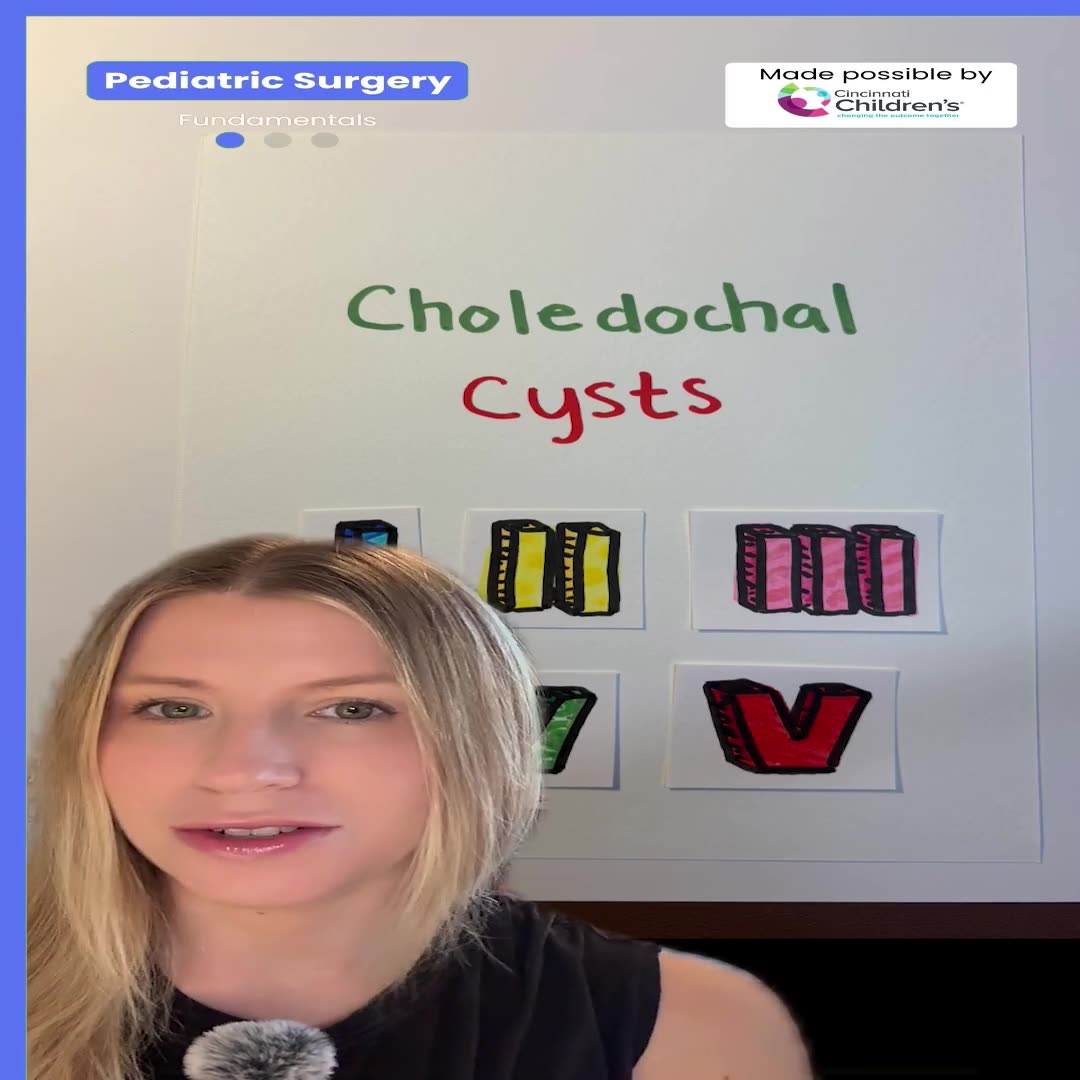
Choledochal Cyst Types Explained: Pediatric Surgery Fundamentals
1 min · Published Sep 2026
What the experts said
The 5 millimeter cool seal with its fine dissecting tips and dual action jaws allows for excellent dissection around delicate structures.
In smaller patients, the surgeon will often take the pulmonary artery vessel at the segmental level, using the cool seal to seal the vessel and safely divide it.
In a larger patient, it is often more efficient to find the pulmonary artery to the lower lobe as it crosses through the fissure, allowing the surgeon to get the main trunk above the bifurcation to the apical and basal segments and use a vascular stapler to safely take the vessel, saving time compared to dissecting into the lung to find the segmental vessels.
In many cases, the surgeon will take the bronchus next after the pulmonary artery as it presents itself before taking the inferior pulmonary vein.
In this case, because of the large tumor and the large amount of inflammation in this area, dissection of the bronchus was difficult, and the surgeon did not want to injure the vein in dissecting this out, so approached the inferior pulmonary ligament instead.
The patient was a 20-year-old who had previously been treated for osteosarcoma in the limb with a limb preservation and had been disease-free for 4 years when she developed shortness of breath and eventually hemoptysis.
CT scan showed a large mass in the left lower lobe.
Three ports were used for the procedure initially 3 5-millimeter ports, with 1 port being increased to a 12 for the stapler.
The 5 millimeter Trinity was the main instrument used for the entire procedure.
The major fissure was relatively complete, however there was still lung tissue overlying the pulmonary artery.
The inferior pulmonary vein was divided a good 1 centimeter away from the pericardium to allow for safe division.
The entire procedure was done in 90 minutes through 2 5-millimeter and 1 12-millimeter trochar.
The patient recovered well and was discharged on the second postoperative day.