

Educational content from recorded physician discussions — not medical advice. Talk to your (or your child's) care team about your situation.
Video
sealer demo
Dr. Steve Rothenberg · 3 min · Published May 2026
Video
pyloric knife
Dr. Steve Rothenberg · Published May 2026
Video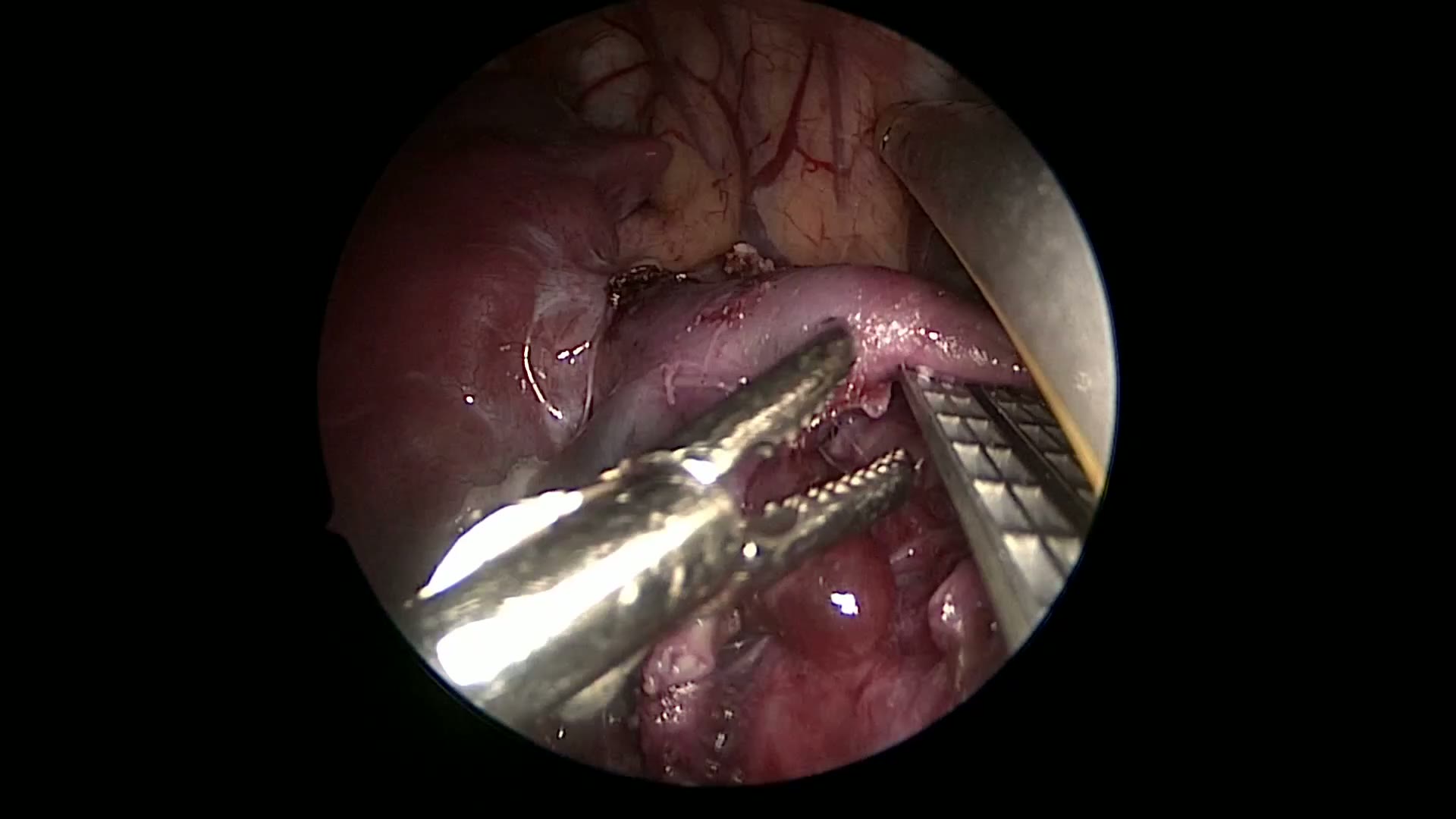
pulm artery LLL
Dr. Steve Rothenberg · 9 s · Published May 2026
Video
plication narrated
Dr. Steve Rothenberg · 6 min · Published May 2026
Video
percuvance redo nissen
Dr. Steve Rothenberg · 5 min · Published May 2026
Video
open bowel anast
Dr. Steve Rothenberg · Published May 2026
Video
Intestinal Atresia Types Explained: Grossfeld Classification for Pediatric Surgery
1 min · Published Sep 2026
Video
Derivation and validation of the Pediatric Community-Acquired Pneumonia Severity (PedCAPS) score: A prospective cohort study
50 s · Published Sep 2026
Video
FETO for Late-Diagnosed Severe Congenital Diaphragmatic Hernia (CDH) at Cincinnati Children's with Dr. Beth Rymeski
4 min · Published Sep 2026
Video
Severe Congenital Diaphragmatic Hernia (CDH) Case: FETO Management & Unexpected Findings with Dr. Beth Rymeski
3 min · Published Sep 2026
Video
Fetoscopic Endoluminal Tracheal Occlusion (FETO) Procedure: Step-by-Step Guide with Dr. Beth Rymeski
4 min · Published Sep 2026
Video
Choledochal Cyst Types Explained: Pediatric Surgery Fundamentals
1 min · Published Sep 2026
What the experts said
Prenatal imaging showed a 4.5 by 4 by 4 centimeter left lower lobe mass
Chest X-ray at birth showed diffuse hazy lungs with no discrete masses
CT at 3 months of age showed a dense focal opacity in the left lower lobe with feeding vessel arising from the celiac axis below the diaphragm and branching within the sequestration
Several cysts at the periphery of the lesion were noted on CT imaging
An anterior approach was used using three ports: a 4 millimeter through the posterior axillary line for the telescope, two 3 millimeter ports placed in the anterior axillary line
The lower port was later changed to a 5 millimeter port for access of the 5 millimeter stapler and 5 millimeter clip applier
Several enlarged lymph nodes were present within the inferior pulmonary ligament
Adjacent to the artery was a cystic structure from which mucinous material was expressed
The systemic artery was double clipped on both sides and then divided with 3 millimeter scissors
An incomplete major fissure was identified and opened using a combination of 5 millimeter stapler, 3 millimeter scissors, and 3 millimeter sealer device
The major fissure was completed in a plane that was superficial to the pulmonary artery branches
The 3 millimeter sealer was first applied to the tissue to allow for cautery as well as crushing of the tissue prior to applying the 3 millimeter scissors
Each of the two segmental bronchi was divided using the 5 millimeter stapler
Placement of the Maryland clamp at the base of the vessel prior to firing of the stapler provides a protective measure in case the staples do not incorporate the entire line of transaction
The lowest incision was expanded from 5 millimeters to 1 centimeter, and the specimen removed piecemeal in its entirety
The chest tube was removed on post-op day 1 and the patient discharged home on post-op day 2
Final pathology showed lung lobe with sequestration and secondary changes of cystic pulmonary airway malformation
Early thoracoscopic surgery can be safely performed in infants with complex congenital lung malformations
The 3 millimeter sealer and 5 millimeter stapler facilitate a thorough dissection and complete excision of tissue in complex congenital lung malformations
Early surgical approach promotes conservation of lung parenchyma and reduces postoperative thoracic musculoskeletal deformity