

Educational content from recorded physician discussions — not medical advice. Talk to your (or your child's) care team about your situation.
Video
sealer demo
Dr. Steve Rothenberg · 3 min · Published May 2026
Video
pyloric knife
Dr. Steve Rothenberg · Published May 2026
Video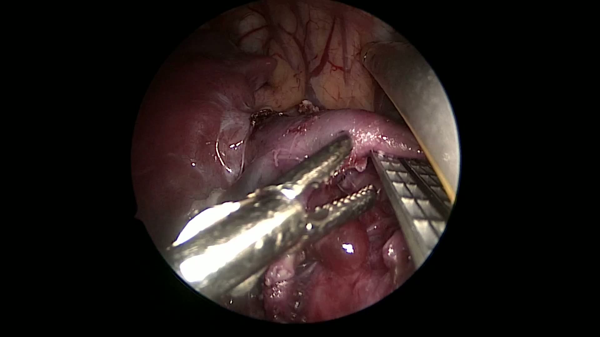
pulm artery LLL
Dr. Steve Rothenberg · 9 s · Published May 2026
Video
plication narrated
Dr. Steve Rothenberg · 6 min · Published May 2026
Video
percuvance redo nissen
Dr. Steve Rothenberg · 5 min · Published May 2026
Video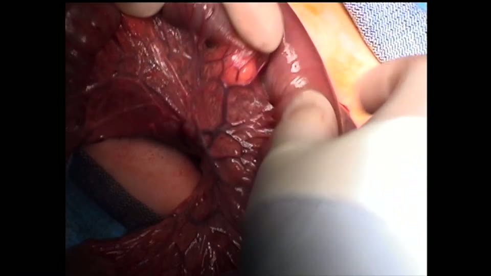
open bowel anast
Dr. Steve Rothenberg · Published May 2026
Video
Intestinal Atresia Types Explained: Grossfeld Classification for Pediatric Surgery
1 min · Published Sep 2026
Video
Derivation and validation of the Pediatric Community-Acquired Pneumonia Severity (PedCAPS) score: A prospective cohort study
50 s · Published Sep 2026
Video
FETO for Late-Diagnosed Severe Congenital Diaphragmatic Hernia (CDH) at Cincinnati Children's with Dr. Beth Rymeski
4 min · Published Sep 2026
Video
Severe Congenital Diaphragmatic Hernia (CDH) Case: FETO Management & Unexpected Findings with Dr. Beth Rymeski
3 min · Published Sep 2026
Video
Fetoscopic Endoluminal Tracheal Occlusion (FETO) Procedure: Step-by-Step Guide with Dr. Beth Rymeski
4 min · Published Sep 2026
Video
Choledochal Cyst Types Explained: Pediatric Surgery Fundamentals
1 min · Published Sep 2026
What the experts said
The patient is a 16-year-old female who presented with a 24 hour history of increasing right lower quadrant pain.
Two ports are placed through a 1 centimeter umbilical incision, and a single 3 millimeter incision is made in the suprapubic area for the left hand retracting port.
The child weighs 65 kg and it is possible to use this technique in patients up to 100 kg.
A 1 centimeter incision is made within the umbilicus so that the scar will not be visible postoperatively.
The abdomen is insufflated to a pressure of 15 mmHg using a closed Veress needle technique in all cases.
A 4 millimeter trochar is inserted for a 4 millimeter 30 degree scope; in larger patients, a 5 millimeter 30 degree scope is often used.
A second trochar is inserted through the same incision but through a separate fascial defect for the 5 millimeter Maryland, hook cautery, and eventually the 5 millimeter stapler.
A 3 millimeter incision is made just above the pubic bone, below the pubic hairline, for a 3 millimeter Babcock which provides right angle retraction.
The appendix was mildly inflamed.
Hook cautery is used to take the appendiceal mesentery; there is significant energy spread and smoke, but this is acceptable because the appendix is being removed.
Monopolar cautery is used in this case; the dissection can also be done using a 3 millimeter sealer or other energy devices, but a reusable hook cautery is generally adequate.
It is important to not leave the appendiceal artery patent next to the appendix when using the 5 millimeter stapler, as sometimes the vessel is so small the staples do not adequately capture it.
In patients with a thicker mesentery, the staple load can be used for the mesentery, although the hook cautery technique works quite well.
The base of the appendix is grasped and compressed to ensure it is not too thick for the 5 millimeter stapler.
The 5 millimeter trocar is removed and a 5 millimeter endoscopic bag is inserted through the fascial defect, opened in the abdominal cavity, and the appendix is placed within it.
The 5 millimeter stapler is compressed with checking to ensure excellent alignment of the two ends; if necessary, the stapler can be reapplied.
There is occasionally some very mild oozing at the staple line, which is insignificant.
The bag is removed through the umbilicus; if necessary, the fascial defect is widened or the two fascial defects are joined using a Kelly clamp, but the patient remains with a 1 centimeter skin incision.