

From
StayCurrentMD
Thoracoscopic Right Upper Lobectomy in a 3-Month-Old with Incomplete Fissures
With Dr. Steve Rothenberg
Educational content from recorded physician discussions — not medical advice. Talk to your (or your child's) care team about your situation.
Video
sealer demo
Dr. Steve Rothenberg · 3 min · Published May 2026
Video
pyloric knife
Dr. Steve Rothenberg · Published May 2026
Video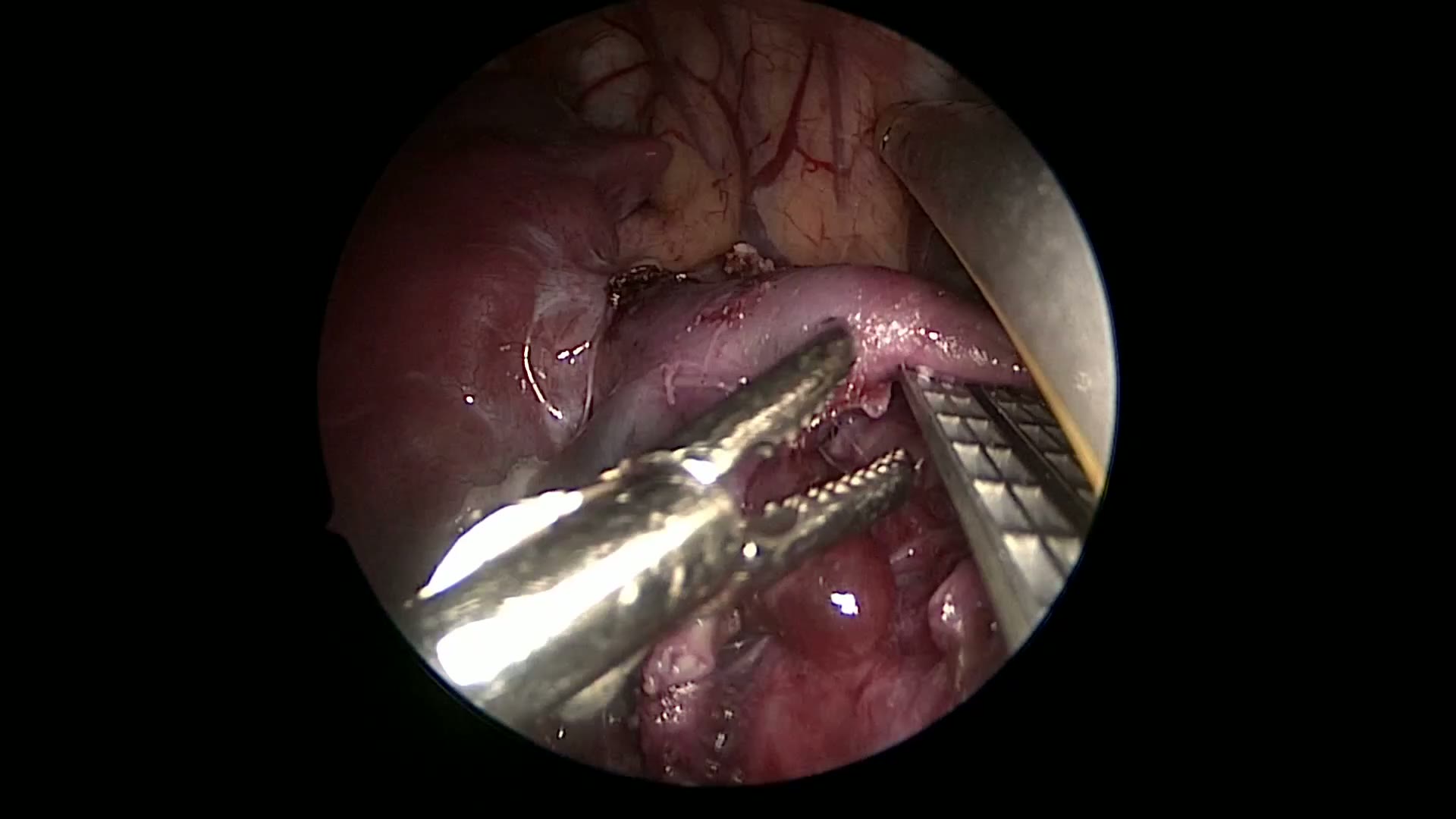
pulm artery LLL
Dr. Steve Rothenberg · 9 s · Published May 2026
Video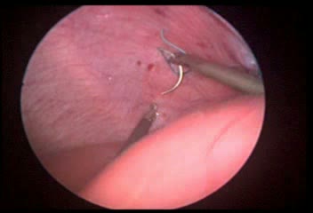
plication narrated
Dr. Steve Rothenberg · 6 min · Published May 2026
Video
percuvance redo nissen
Dr. Steve Rothenberg · 5 min · Published May 2026
Video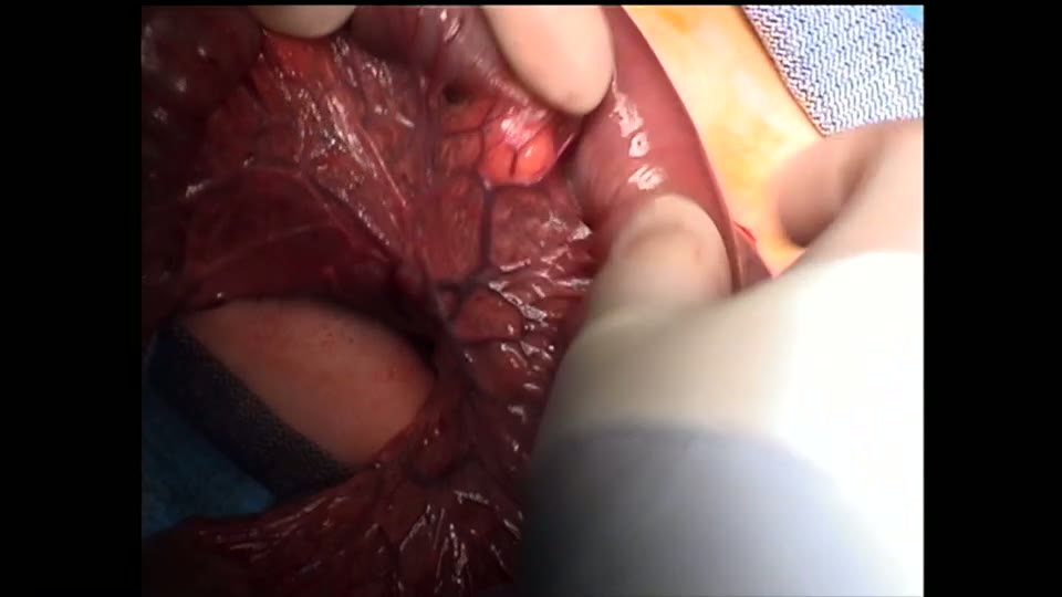
open bowel anast
Dr. Steve Rothenberg · Published May 2026
Video
Intestinal Atresia Types Explained: Grossfeld Classification for Pediatric Surgery
1 min · Published Sep 2026
Video
Derivation and validation of the Pediatric Community-Acquired Pneumonia Severity (PedCAPS) score: A prospective cohort study
50 s · Published Sep 2026
Video
FETO for Late-Diagnosed Severe Congenital Diaphragmatic Hernia (CDH) at Cincinnati Children's with Dr. Beth Rymeski
4 min · Published Sep 2026
Video
Severe Congenital Diaphragmatic Hernia (CDH) Case: FETO Management & Unexpected Findings with Dr. Beth Rymeski
3 min · Published Sep 2026
Video
Fetoscopic Endoluminal Tracheal Occlusion (FETO) Procedure: Step-by-Step Guide with Dr. Beth Rymeski
4 min · Published Sep 2026
Video
Choledochal Cyst Types Explained: Pediatric Surgery Fundamentals
1 min · Published Sep 2026
What the experts said
The patient is a 3-month-old with type 2 CPAM with significantly incomplete fissures undergoing thoracoscopic right upper lobectomy.
Patient positioning uses left lateral decubitus position with surgeon and assistant standing at the patient's front and monitors placed posteriorly.
The scope is placed in approximately the 5th intercostal space just posterior to the mid-axillary line, with right and left operating ports placed above and below in the anterior axillary line.
Initial port sizes are 4mm trocar for the scope and two 3mm ports for operating, with a 3mm vessel sealer used as the primary dissection mode for the entire procedure.
Dissection starts at the top where the pulmonary artery transverses below the upper lobe to devascularize the upper lobe and decrease congestion during the rest of the dissection.
Vessels are individually isolated and sealed proximally and distally with at least 3-4mm between the seals before division, providing the safest approach to pulmonary vasculature.
Making two separate seals and then dividing between them ensures the vessel is adequately sealed before it retracts, preventing bleeding that could lead to conversion to open.
If inadequate length of vessel cannot be obtained for adequate vascular control, one should dissect down into the lung parenchyma to create length.
The vessel sealer takes vessels up to 5mm in diameter; most subsegmental vessels at this level in this size child are 2-3mm in diameter.
For the upper lobe, it is often necessary to take vessels at the subsegmental level to ensure no injury to vessels transversing behind the upper lobe and going to the middle and lower lobe, as opposed to the lower lobe where the main trunk can easily be identified.
In this case, the major fissure is complete anteriorly but the minor fissure is almost non-existent, with only a small cleft posteriorly where the minor fissure adjoins the major fissure.
The sealer is an excellent way to go across lung parenchyma in the area of incomplete fissure, developing the fissure in a layer-by-layer process by sealing and then dividing in the middle of the seal.
Compressing the upper lobe using the energy device works extremely well with large cysts but even works when the cysts are much smaller, improving visualization of the pulmonary vein.
Care must be taken when dividing the superior pulmonary vein to ensure there is no injury to the vein to the middle lobe.
There is usually a clear delineation between the middle lobe vein and the upper lobe veins, but extreme care must be taken to preserve the middle lobe.
With the vein to the upper lobe completely divided, it helps differentiate between the upper and middle lobe, and the ischemic upper lobe can be used along with the posterior congenital cleft to guide fissure creation.
Creating the fissure layer-by-layer with the sealer is very similar to finger fracturing of the liver during a hepatic segmentectomy.
There is minimal bleeding during fissure creation because of the devascularization of the upper lobe and because the fissure is a relatively avascular plane.
This method of fissure creation is very hemostatic and results in no air leak or significant air leak postoperatively.
In cases of complete fissure, the dissection portion is much quicker, requiring only a few minutes rather than 10-15 minutes.
There is usually a branch of the artery coming back up from the pulmonary artery to the anterior segmental segment that should be sealed and divided.
The upper lobe bronchus is dissected past the point of bifurcation, taking the apical and posterior segmental bronchus separately from the anterior segmental bronchus.
The lower port is changed to a 5mm port to allow insertion of a 5mm endoscopic stapler for bronchial division.
The stapler can also be used to complete the incomplete major fissure posteriorly.
The lung is removed piecemeal through an enlarged incision site using a tonsil clamp.
This operation took 1 hour and 35 minutes.