

From
StayCurrentMD
Sacral Nerve Stimulator Placement: A Novel Surgical Navigation Tool for...
With CCHMC Pediatric Surgery
Educational content from recorded physician discussions — not medical advice. Talk to your (or your child's) care team about your situation.
Video
What is Hirschsprung's Disease? An ERNICA animation for parents and families
4 min · Published Dec 2023
Video
Bowel Management - Bowel Management In Communities With Limited Resources
9 min · Published Nov 2018
Video
Enterocolitis in Hirschsprung Disease: Update Course 2015
CCHMC Pediatric Surgery · 5 min · Published Nov 2015
Video
Surgical treatment for Hirschsprung’s Disease: An ERNICA animation for parents and families
3 min · Published Dec 2023
Video
Use of hydrocolonic sonography to monitor the effects of an enema: Pediatric...
Dr. Todd Ponsky · 12 min · Published May 2013
Video
Bowel Management - Use Of Ultrasound To Monitor Effects Of Enema
12 min · Published Nov 2018
Video
Intestinal Atresia Types Explained: Grossfeld Classification for Pediatric Surgery
1 min · Published Sep 2026
Video
Derivation and validation of the Pediatric Community-Acquired Pneumonia Severity (PedCAPS) score: A prospective cohort study
50 s · Published Sep 2026
Video
FETO for Late-Diagnosed Severe Congenital Diaphragmatic Hernia (CDH) at Cincinnati Children's with Dr. Beth Rymeski
4 min · Published Sep 2026
Video
Severe Congenital Diaphragmatic Hernia (CDH) Case: FETO Management & Unexpected Findings with Dr. Beth Rymeski
3 min · Published Sep 2026
Video
Fetoscopic Endoluminal Tracheal Occlusion (FETO) Procedure: Step-by-Step Guide with Dr. Beth Rymeski
4 min · Published Sep 2026
Video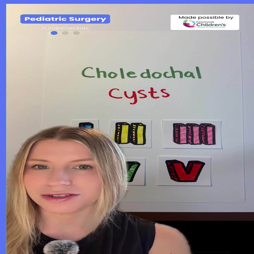
Choledochal Cyst Types Explained: Pediatric Surgery Fundamentals
1 min · Published Sep 2026
What the experts said
Placement in our institution happens in a hybrid operating room equipped with a C-ARM cone beam CT and a surgical navigation system.
Using this technique enables us to offer this therapy to patients with dysplastic or malformed sacra whose anatomy would be otherwise prohibitive for traditional or purely fluoroscopic interventions.
The patient is placed under general anesthesia and position prone with a pelvic roll for elevation and stabilization.
The horizontal axis is marked at the posterior inferior iliac spine in the cranial aspect of the sciatic notch.
The vertical axis are traced overlying the medial aspect of the S3 neural foramina bilaterally, roughly 2 centimeters from the midline.
Conventional skin entry points are marked 2 centimeters cranially to these intersections.
A cone beam CT run is acquired intraoperatively and a three dimensional model constructed for procedural guidance.
The planned needle entry site was identified around 1 centimeter caudal to the traditional markings in this case.
A bellows contraction identifies proper placement at the S3 foramen with confirmation by plantar flexion of the great toe.
The marker should be visible halfway through the foramen.
A tined lead is then inserted under live visualization until the 3rd and 4th marks straddle the interior edge of the sacrum.
This lead consists of 4 distal electrodes, tines or barbs for stabilization, distance markers, and 4 proximal connectors.
Over the next 2 to 4 weeks, the patient's bowel function is carefully recorded and compared to previous records.
If a positive effect is demonstrated with neuromodulation, a permanent generator is implanted in a second stage.
If unsuccessful, the lead can be removed without permanent damage to the nerve.
Sacral neuromodulation is a therapy aimed at improving the quality of life of patients with constipation and fecal incontinence by delivering electrical impulses through a probe placed near the sacral nerve.
This technology acts like a pacemaker to promote bowel and sphincter function.
These would provide access to the S3 foramina at a 60 degree angle in the traditional approach without navigation.