

From
StayCurrentMD
Esophageal Atresia in Brief: Presentation, Workup, Diagnosis, and Treatment
With Dr. Daniel von Allmen & Dr. Aaron Garrison
Educational content from recorded physician discussions — not medical advice. Talk to your (or your child's) care team about your situation.
Video
Thoracoscopic TEF Repair, Is It Really Better Than Open? Update Course 2018
18 min · Published Aug 2018
Video
Update Course 2013: EA & TEF
38 min · Published Sep 2013
Video
EA/TEF Discussion & Technique: Difficult Cases
19 min · Published Dec 2013
Video
Leak after Esophageal Atresia Repair / TEF Repair: Discussion of World Experts
Dr. Todd Ponsky · 15 min · Published Dec 2012
Video
Thoracoscopic Repair of Tracheo-esophageal Fistula Tricks: Pediatric Surgery...
19 min · Published Jul 2017
Video
QUAD #10 Pt.3: Advantages of Thoracoscopic Repair of Esophageal Atresia with Dr. Steve Rothenberg
Dr. Steve Rothenberg · Published Jun 2024
Podcast
Treatment for Long Gap Esophageal Atresia: Esophageal Elongation and Replacement
10 min · Published Jan 2022
Video
7th Annual Pediatric Surgery Update Course 2019 - FULL SHOW
285 min · Published Jul 2020
Podcast
Neuroblastoma
56 min · Published Aug 2019
Video
Aerodigestive & Esophageal Surgery - The Unsalvageable Esophagus & Cases
CCHMC Pediatric Surgery · 101 min · Published Nov 2018
Video
Aerodigestive & Esophageal Surgery - Difficult Tracheal Esophageal Fistula
Dr. Todd Ponsky · 120 min · Published Nov 2018
Video
Abdominal Wall Defects
37 min · Published Nov 2018
Video
Intestinal Atresia Types Explained: Grossfeld Classification for Pediatric Surgery
1 min · Published Sep 2026
Video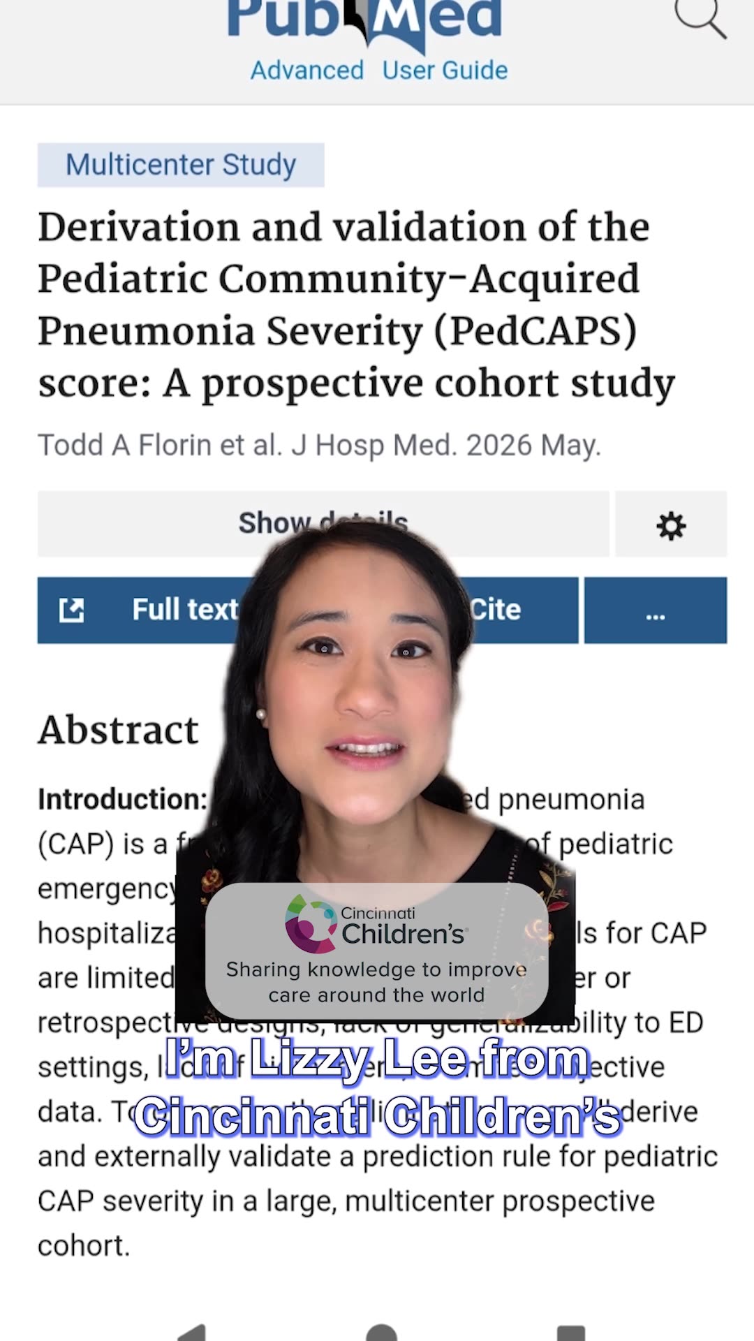
Derivation and validation of the Pediatric Community-Acquired Pneumonia Severity (PedCAPS) score: A prospective cohort study
50 s · Published Sep 2026
Video
FETO for Late-Diagnosed Severe Congenital Diaphragmatic Hernia (CDH) at Cincinnati Children's with Dr. Beth Rymeski
4 min · Published Sep 2026
Video
Severe Congenital Diaphragmatic Hernia (CDH) Case: FETO Management & Unexpected Findings with Dr. Beth Rymeski
3 min · Published Sep 2026
Video
Fetoscopic Endoluminal Tracheal Occlusion (FETO) Procedure: Step-by-Step Guide with Dr. Beth Rymeski
4 min · Published Sep 2026
Video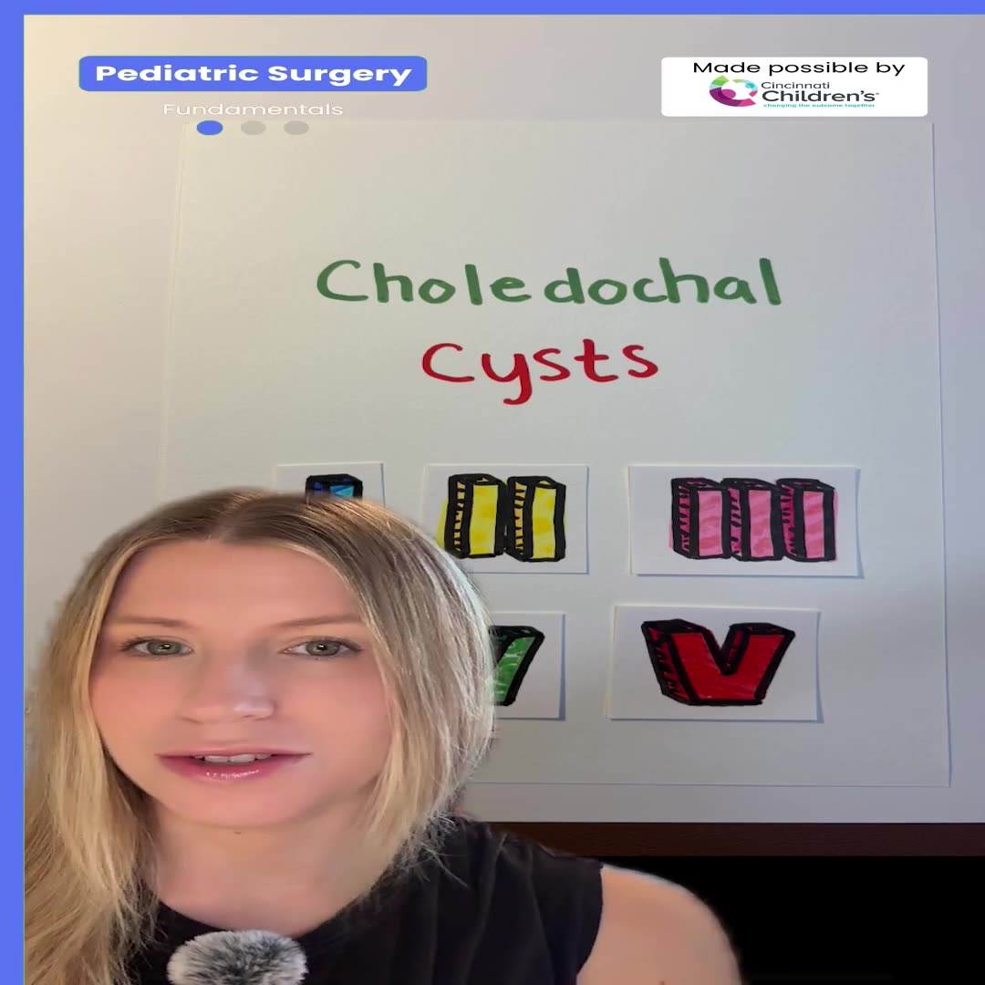
Choledochal Cyst Types Explained: Pediatric Surgery Fundamentals
1 min · Published Sep 2026
What the experts said
Esophageal atresia affects 1 in 2,500 live births.
Esophageal atresia is a congenital anomaly in which the esophagus doesn't form completely, with multiple different variants, one of the most common including a tracheoesophageal fistula.
Infants with isolated esophageal atresia are sometimes diagnosed prenatally because the mother can have polyhydramnios during gestation.
Patients with tracheoesophageal fistula usually don't present until after birth when they have problems feeding, and a Repogle tube meets resistance at typically 10 to 12 centimeters.
X-ray after NG tube placement demonstrates the tube coiled in the upper part of the chest in the proximal pouch of the atretic esophagus.
Injecting air into the NG tube prior to X-ray will distend the proximal pouch; if it doesn't distend, either the diagnosis is wrong or there could be a proximal fistula.
40 to 60% of babies with esophageal atresia can have other associated anomalies.
About 20% of babies with esophageal atresia can have other findings within the VACTERL association.
About 5% of babies with esophageal atresia can have genetic syndromes.
The VACTERL association includes vertebral anomalies, anorectal malformations, cardiac anomalies, tracheoesophageal fistula, renal defects, and limb anomalies.
The most pressing study needed is an echocardiogram to look at cardiac function and ensure there are no other congenital heart anomalies, and to identify which side the aortic arch is on.
VACTERL workup includes renal ultrasound and spine ultrasound, which doesn't need to be done immediately after birth.
To be considered truly VACTERL, you need at least three of the associations.
There are five different types of EA or TEF, with two types accounting for over 90% of presentations.
Type C is the most common type of esophageal atresia, which is esophageal atresia with a distal fistula where the esophagus distally fistulizes to the trachea, usually a couple centimeters above the carina.
Type A (isolated esophageal atresia with no fistula to the trachea) is the next most common type at about 8% of incidence.
H-type (more accurately N-type) esophageal atresia has an intact esophagus but a fistula between the trachea and esophagus, typically high, and is uncommon.
Other very uncommon types include proximal fistula or proximal and distal fistula to the trachea.
Initial management involves ensuring the Repogle is in a good position to decompress saliva or secretions and minimize aspiration while completing the workup.
For the most common type of esophageal atresia, management is to stabilize the patient and get them to the OR, with almost all babies needing surgery within the first day or two depending on stability and medical comorbidities.
Every child with esophageal atresia should undergo bronchoscopy at their initial trip to the operating room.
Bronchoscopy is performed to evaluate for the location of a proximal fistula, evaluate the degree of tracheomalacia, and help position the endotracheal tube to minimize the risk of insufflating the GI tract.
Type C esophageal atresia can be repaired thoracoscopically or open, with the operation involving ligation of the tracheoesophageal fistula and bringing the two ends of the esophagus together to create a new anastomosis.
Chest tubes are almost always used after repair.
Extubation is attempted in one to two days, minimizing the risk of using high flow or CPAP following extubation, so the child should be in good respiratory shape when the tube is pulled.
Most surgeons get an esophagram five to seven days after the operation to check for leak prior to removing the chest tube or starting feeds.
One of the most common short-term complications is a leak following repair, usually diagnosed with spit coming out of the chest tube, and managed with observation.
The second most common complication is stricture.
Recurrent fistula is a less common early complication.
Reflux is a longer-term complication.
Other long-term complications include respiratory issues like reactive airway disease and esophageal dysmotility.
Survival of esophageal atresia patients has improved over the years and is now about 85 to 95% overall survival.
Lower survival is typically seen in more complex patients with other cardiac, renal, and/or pulmonary anomalies.
Having a multidisciplinary approach that's coordinated is important to achieving the best outcomes for these patients in the long term.