

From
Grand Rounds
Ovarian Tumors with Dr. Roshni Dasgupta
With Dr. Roshni Dasgupta · hosted by Dr. Em Gootee or Todd Ponsky
Educational content from recorded physician discussions — not medical advice. Talk to your (or your child's) care team about your situation.
Video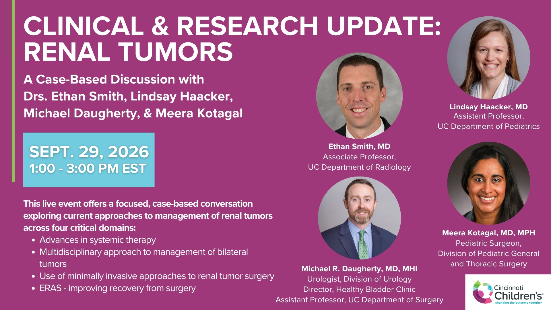
Clinical & Research Update: Renal Tumors with Drs. Ethan Smith, Lindsay Haacker, Michael Daugherty, and Meera Kotagal
71 min · Published Sep 2026
Video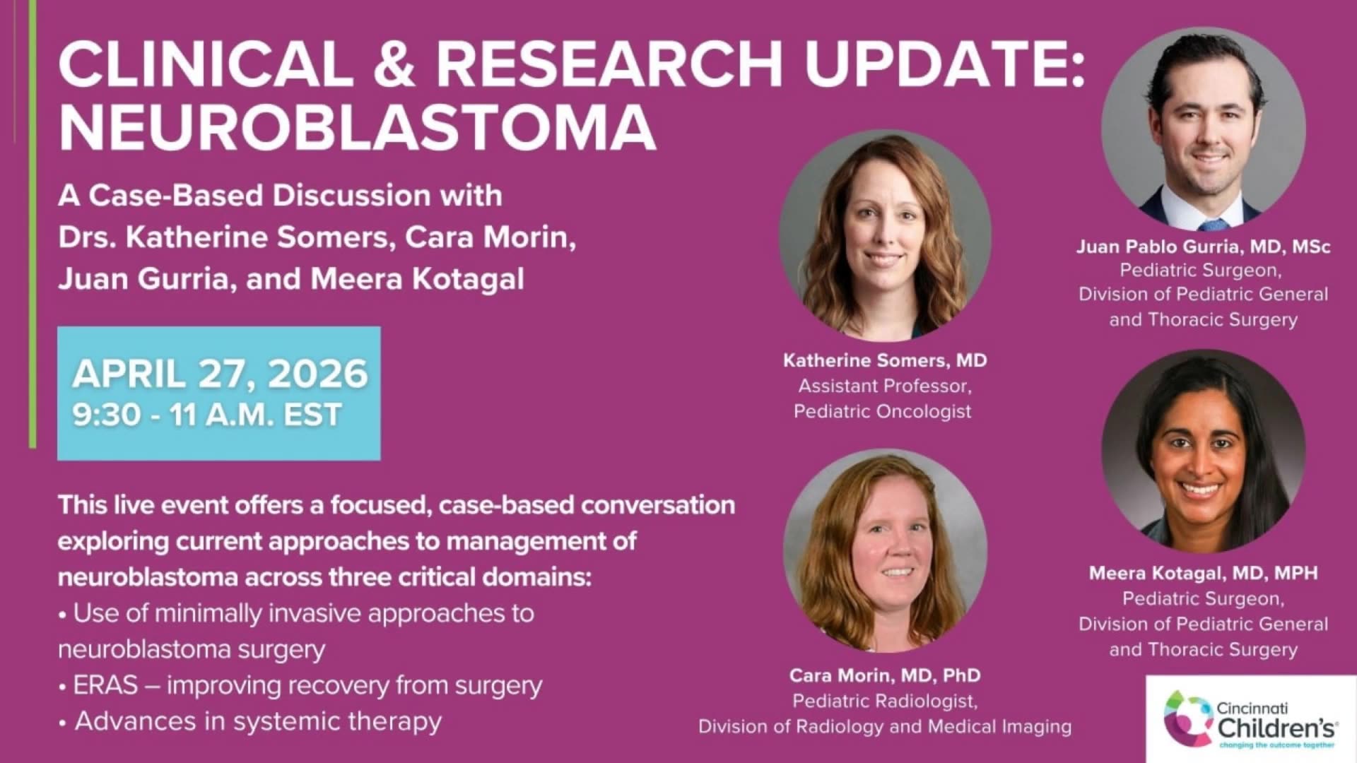
Clinical & Research Update: Neuroblastoma with Drs. Katherine Somers, Cara Morin, Juan Gurria, and Meera Kotagal
67 min · Published Sep 2026
Video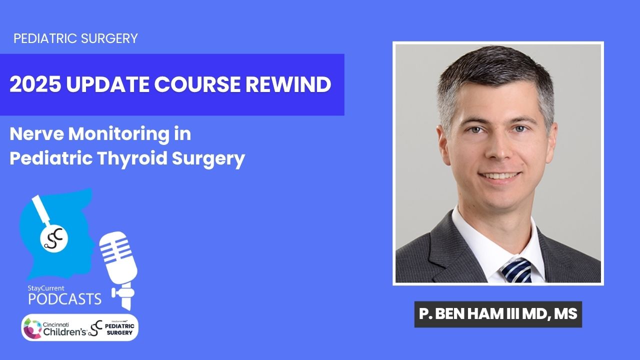
Update Course Rewind 2025: Nerve Monitoring in Pediatric Thyroid Surgery
6 min · Published Jul 2026
Video
Journal of Pediatric Surgery Article Review: June 2023, AAP Issue
Todd Ponsky · 13 min · Published Jul 2026
Video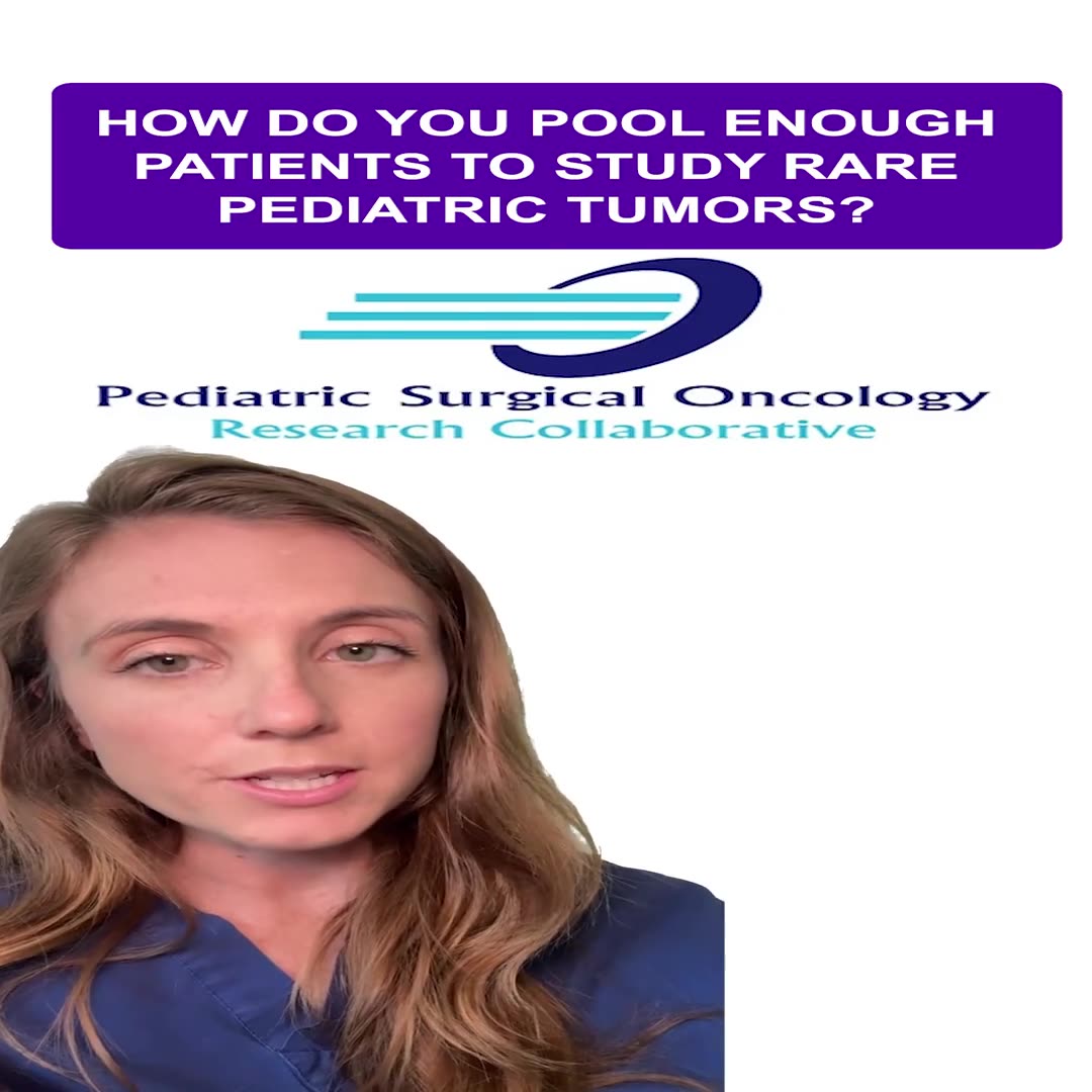
Pediatric Surgical Oncology Research Collaborative (PSORC): Studying Rare Pediatric Tumors
56 s · Published May 2026
Video
Pooling Patients to Study Rare Pediatric Tumors: An Introduction to PSORC
56 s · Published May 2026
Video
Clinical & Research Update: Sarcoma w/ Drs. Roshni Dasgupta, Joseph Pressey, Arthur Meyer, Luke Pater
66 min · Published Sep 2026
Video
Ovarian Torsion with Dr. Dasgupta
Published Sep 2022
Video
Ovarian Cysts with Dr. Roshni Dasgupta
Published Aug 2022
Video
Update Course Rewind: Thoracotomy vs VATS for Lung Metastases
Dr. Todd Ponsky · 9 min · Published Dec 2021
Video
GYN #4 Management of Tubal Torsion: When to Consider Salpingectomy with Dr. Lesley Breech
CCHMC Pediatric Surgery · Published Oct 2024
Podcast
Pyloric Stenosis with Dr. Alex Bondoc
16 min · Published Jun 2024
Video
GYN #3 Management of an Adnexal Torsion with a Healthy Appearing Ovary with Dr. Lesley Breech
CCHMC Pediatric Surgery · Published May 2024
Video
GYN #2 Oophoropexy in Adnexal Torsion with Dr. Lesley Breech
CCHMC Pediatric Surgery · Published May 2024
Video
GYN #1 Importance of Documenting Reproductive Anatomy with Dr. Lesley Breech
CCHMC Pediatric Surgery · Published Feb 2024
Podcast
Esophageal Replacement with Dr. Dan von Allmen
13 min · Published Nov 2023
What the experts said
Neoplastic ovarian masses are classified into three types based on tissue of origin: epithelial ovarian tumors, stromal/sex cord tumors, and germ cell tumors (from the yolk sac).
Malignancy from neoplastic ovarian processes is rare, making up only 2% of cancers in children overall.
Ovarian malignancy is more common in the 10-19 year age group and any malignant lesion under age 5 is reportable.
Peutz-Jeghers syndrome is associated primarily with granulosa cell tumors and sex cord tumors.
Ollier and Maffucci syndromes are associated with ovarian tumors.
Chediak-Higashi and McCune-Albright syndromes are also associated with ovarian tumors.
Workup for suspected ovarian tumor with precocious puberty includes renal panel, CBC, estradiol, progesterone, tumor markers, and ultrasound.
CT scan advantages for ovarian mass evaluation include better determination of tumor location and extent, and superior detection of calcifications compared to MRI.
CT scan disadvantage is radiation exposure to the child.
MRI provides better tissue detail, can help distinguish ovarian from parovarian masses, and is excellent at detecting lymph nodes.
Tumor markers commonly associated with ovarian tumors include AFP, beta-hCG, CA-125 (for epithelial tumors), inhibin A and B, hormones (estradiol, progesterone), LDH, CBC, and renal function tests.
Elevated inhibin B is associated with granulosa cell tumor, a sex cord tumor that causes virilizing effects.
CA-125 can be elevated in benign conditions such as endometriosis and other inflammatory conditions, sometimes to levels that overlap with malignancy.
Primordial germ cells form in the first week of gestation, and by week three they form the wall of the yolk sac and migrate along the mesentery of the hindgut.
Germ cell tumors can be gonadal (when germ cells migrate to the gonads) or extragonadal (sacrococcygeal teratomas, mediastinal lesions).
Within gonadal germ cell tumors, if cells differentiate properly, extraembryonic differentiation produces choriocarcinomas and yolk sac tumors, while embryonic differentiation produces teratomas and mixed germ cell tumors.
If germ cells do not differentiate properly, they are classified as seminomas in boys and dysgerminomas in girls.
About 20% of germ cell tumors are malignant.
Dysgerminomas are the most common malignant histology, comprising 30% of germ cell tumors, and can be bilateral.
Dysgerminomas have a unique laboratory pattern: elevated LDH, but in a pure dysgerminoma AFP and beta-hCG can be normal; hypercalcemia may also be present.
Germ cell tumor staging requires peritoneal washings, examination of the entire peritoneal cavity, omental resection only if suspicious nodules are present, and unilateral salpingo-oophorectomy (salpingectomy only if fallopian tube is involved).
For germ cell tumors, lymph nodes should be examined intraoperatively but only removed if enlarged or suspicious; standard lymph node dissection is not required.
Any portion of the ovary that can be preserved should be considered during germ cell tumor resection.
COG staging for germ cell tumors: stage I is limited to the ovary with negative washings; stage II includes capsular rupture with negative washings; stage III is lymph node involvement; stage IV is distant metastasis.
Stage I germ cell tumors can be managed with surgery alone and observation if the tumor is not ruptured.
If a germ cell tumor is ruptured during laparoscopic resection and tumor is spilled, the patient is required to receive chemotherapy.
For mature teratomas with a cystic component, it is safe to decompress the cyst first to improve access and increase chances of ovarian preservation.
The ovarian capsule does not need to be closed after shelling out a teratoma; there is no data showing closure is necessary and it will form by itself.
A plastic bowel bag can be glued to the ovarian mass and a needle inserted through it to avoid spillage during teratoma resection.
Spillage of teratoma contents can cause growing teratoma syndrome, where teratoma grows throughout the abdomen and is not responsive to chemotherapy.
Staging for epithelial ovarian tumors requires pelvic washings, abdominal examination, omentectomy, primary tumor removal, and lymph node dissection.
For epithelial tumors, lymph node dissection boundaries are the pelvic iliacs to the renal artery (para-aortic nodes below the renals).
About 30% of normal-appearing lymph nodes in epithelial ovarian cancer are positive on pathology, which will upstage the patient.
Borderline epithelial tumors can recur, so patients require close serial follow-up over many years.
For borderline epithelial tumors, ovarian-sparing surgery can be performed initially, and if pathology confirms borderline tumor, the ovary can either be removed in a second operation or the patient can be watched with close surveillance.
There is no perfect tumor marker for ovarian masses; studies from the Midwest Pediatric Surgery Consortium show that a panel of tumor markers is helpful, but no single marker perfectly predicts malignancy.
MRI disadvantages include longer acquisition time (sometimes requiring anesthesia), reduced availability, and higher cost.
AFP is the primary tumor marker for germ cell disease.
PEB chemotherapy (platinum, etoposide, bleomycin) is the standard approach for germ cell tumors requiring adjuvant treatment and is very effective.
Six-year overall survival with PEB chemotherapy is excellent even in stage IV germ cell tumor patients.
Teratomas consist of all three primordial tissue types (ectoderm, mesoderm, endoderm) and can contain brain, bone, and teeth.
Cystic teratomas are the most common type and can be bilateral in about 10% of patients.
Calcifications are often visible in teratomas, making CT scan useful for detection.
Immature teratomas have neuroepithelial components graded 0-3; higher grades are more likely to have yolk sac foci and malignant behavior.
Most immature teratomas do not need chemotherapy after resection.
Epithelial ovarian tumors account for about 20% of pediatric ovarian tumors, which is relatively rare compared to adults.
Epithelial tumors are mucinous and serous; serous lesions are more commonly bilateral than mucinous.
Borderline epithelial tumors are more common in children and have a better prognosis than invasive ovarian cancer.