

From
StayCurrentMD
Ovarian Teratoma - Ovarian Torsion - Soft Tissue Sarcoma: Update Course 2015
With Dr. Andrea Hayes-Jordan · hosted by Dr. Todd Ponsky
Educational content from recorded physician discussions — not medical advice. Talk to your (or your child's) care team about your situation.
Only a few other public items share this diagnosis — go deeper there →
Video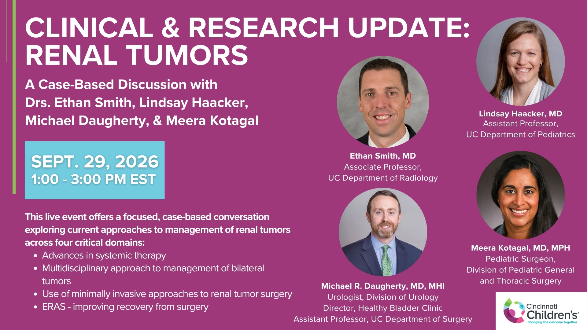
Clinical & Research Update: Renal Tumors with Drs. Ethan Smith, Lindsay Haacker, Michael Daugherty, and Meera Kotagal
71 min · Published Sep 2026
Video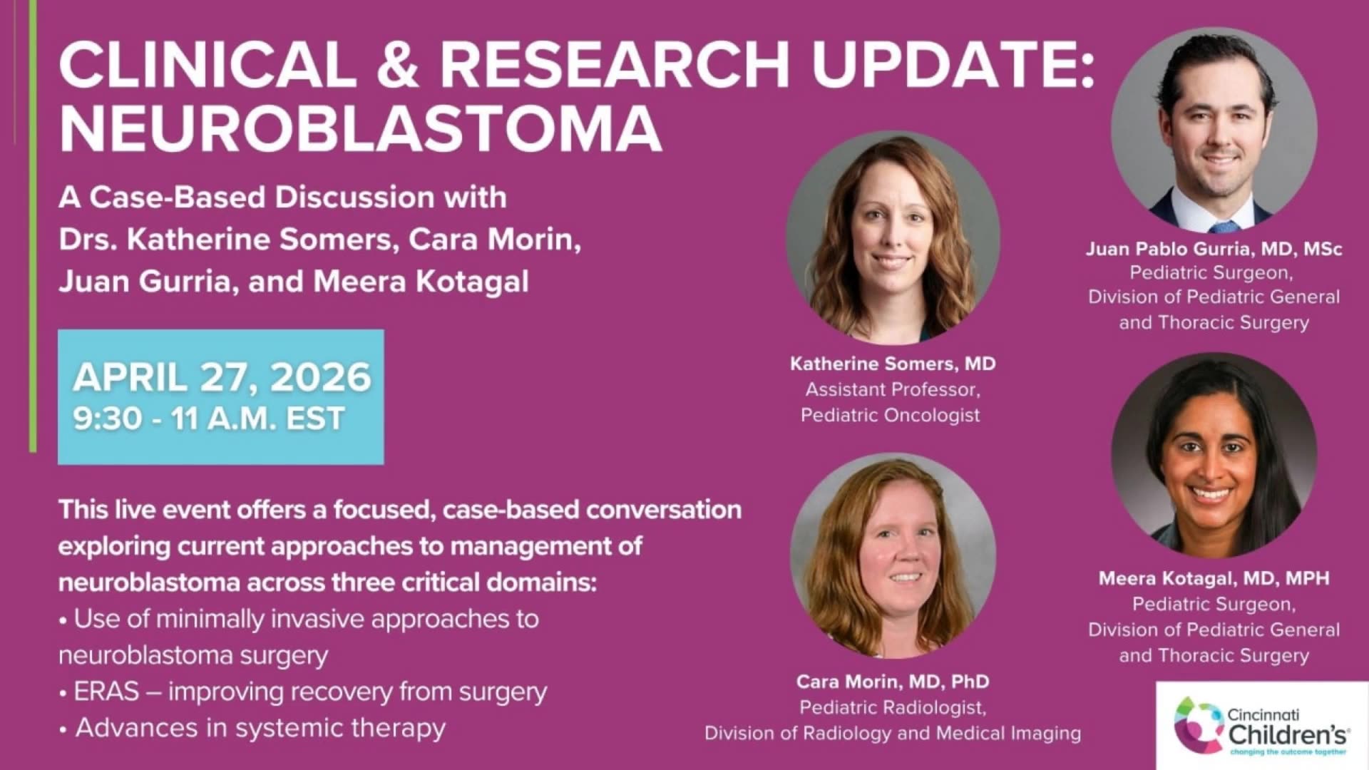
Clinical & Research Update: Neuroblastoma with Drs. Katherine Somers, Cara Morin, Juan Gurria, and Meera Kotagal
67 min · Published Sep 2026
Video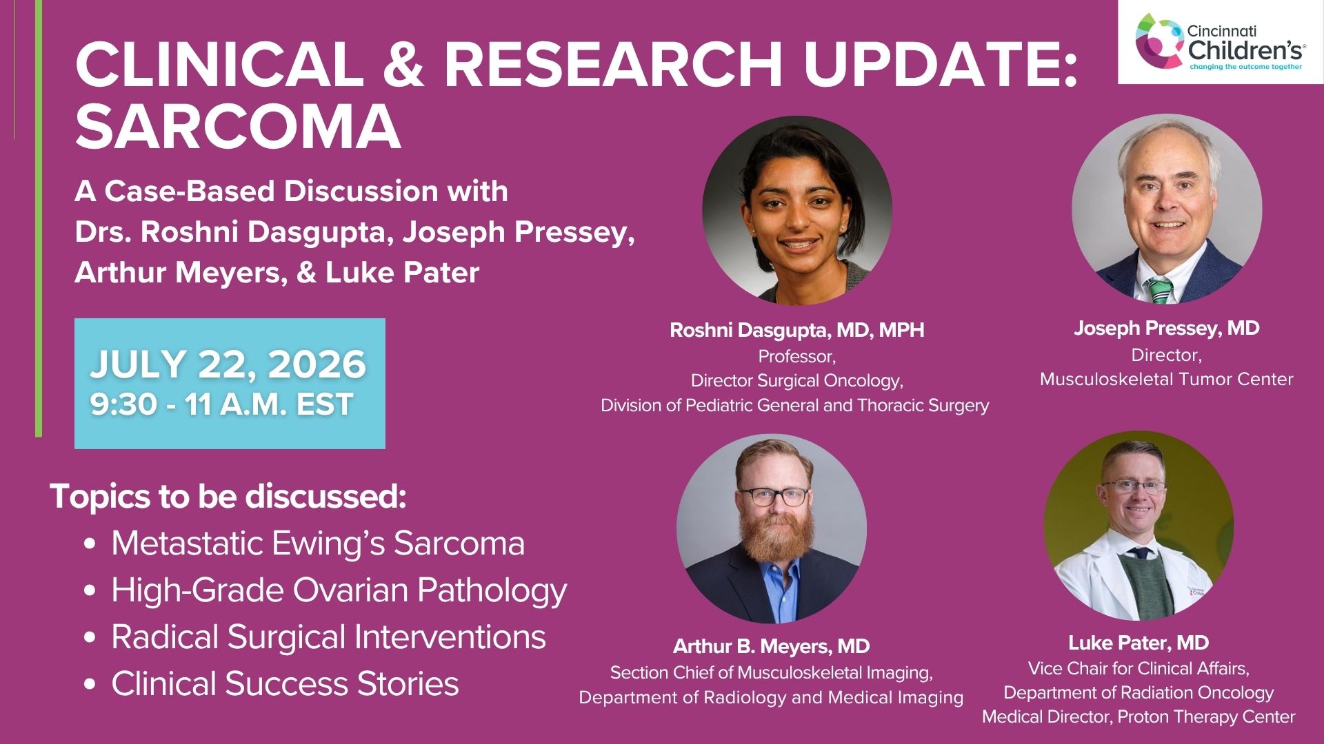
Clinical & Research Update: Sarcoma w/ Drs. Roshni Dasgupta, Joseph Pressey, Arthur Meyer, Luke Pater
66 min · Published Sep 2026
Video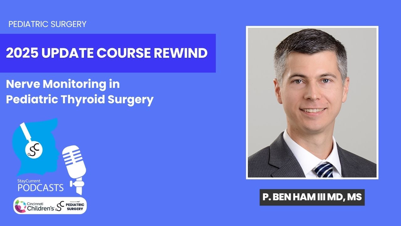
Update Course Rewind 2025: Nerve Monitoring in Pediatric Thyroid Surgery
6 min · Published Jul 2026
Video
Journal of Pediatric Surgery Article Review: June 2023, AAP Issue
Todd Ponsky · 13 min · Published Jul 2026
Video
Pediatric Surgical Oncology Research Collaborative (PSORC): Studying Rare Pediatric Tumors
56 s · Published May 2026
Video
Intestinal Atresia Types Explained: Grossfeld Classification for Pediatric Surgery
1 min · Published Sep 2026
Video
Derivation and validation of the Pediatric Community-Acquired Pneumonia Severity (PedCAPS) score: A prospective cohort study
50 s · Published Sep 2026
Video
FETO for Late-Diagnosed Severe Congenital Diaphragmatic Hernia (CDH) at Cincinnati Children's with Dr. Beth Rymeski
4 min · Published Sep 2026
Video
Severe Congenital Diaphragmatic Hernia (CDH) Case: FETO Management & Unexpected Findings with Dr. Beth Rymeski
3 min · Published Sep 2026
Video
Fetoscopic Endoluminal Tracheal Occlusion (FETO) Procedure: Step-by-Step Guide with Dr. Beth Rymeski
4 min · Published Sep 2026
Video
Choledochal Cyst Types Explained: Pediatric Surgery Fundamentals
1 min · Published Sep 2026
What the experts said
80% of ovarian masses are teratomas
In the year 2000, girls with ovarian germ cell tumors have almost 100% survival
Stage one ovarian tumors limited to the ovary now receive only oophorectomy without chemotherapy
Microscopic residual and lymph node involvement in ovarian tumors require chemotherapy
25% of girls with ovarian germ cell tumors will be missed if peritoneal cytology and washings are not performed
If staging procedures are omitted from the operative report, oncologists will treat ovarian tumors as stage 2
Gliomatosis peritonei is a benign disease appearance associated with ovarian teratomas with nearly 100% survival
In bilateral ovarian disease, tumors larger than 10 cm should definitely be biopsied
If an ovarian tumor is ruptured during laparoscopic removal, the patient is committed to chemotherapy including platinum
15% of ovarian tumors in adolescents are epithelial tumors with different staging criteria than germ cell tumors
If alpha-fetoprotein and beta-HCG levels are normal preoperatively, there is some comfort about draining a cyst without contamination
The salvage rate for recurrent ovarian germ cell tumors is almost 100% because chemotherapy is very effective
Ovarian torsion is an urgent but not emergent situation if the goal is to preserve the ovary
Many black-colored ovaries from torsion have viable follicles and ovarian preservation should be the goal
76% of ovaries removed for torsion had normal ovarian tissue, only 13% had no ovarian tissue, and only 11% were completely necrotic
Pain after oophoropexy is usually short-lived and resolves in about a week
The ability to preserve ovarian function after torsion is related to age, with pediatric patients having more active follicles than older adults
Ultrasound Doppler for ovarian torsion has sensitivity and specificity in the 50-60% range
Half of soft tissue sarcomas are rhabdomyosarcomas and half are non-rhabdo sarcomas
Abdominal, pelvic, and retroperitoneal rhabdomyosarcomas have the worst survival while orbital rhabdomyosarcomas have the best survival
Even small tumors in the extremity or abdomen can only achieve stage 2 at best (not stage 1) in rhabdomyosarcoma staging
Surgical extent determines clinical group in rhabdomyosarcoma: group 1 is complete resection, group 2 is microscopic residual, group 3 is biopsy only
Group 1 and 2 rhabdomyosarcoma patients have excellent prognosis while group 3 patients do more poorly
Inability to resect a large rhabdomyosarcoma is a biologic determination, not a reflection of surgical skill, and aggressive resection with high morbidity will not improve outcome
Sentinel lymph node biopsy is now required for all trunk and extremity rhabdomyosarcomas
For rhabdomyosarcoma, completion node dissection is not performed for positive sentinel nodes; radiation therapy is given instead
Sentinel lymph node mapping for rhabdomyosarcoma should be performed at the time of re-excision if initial excisional biopsy had positive margins
40-50% of biopsied lymph nodes are positive in rhabdomyosarcoma, and clinically negative nodes may also be positive
Histologic grade is now critical in non-rhabdo soft tissue sarcomas, not just the diagnosis
Low-grade soft tissue sarcomas require only resection with observation, no chemotherapy or radiation
Low-grade soft tissue sarcomas with positive margins receive adjuvant radiation therapy
Unresectable soft tissue sarcomas should receive both preoperative radiation and chemotherapy (previously only chemotherapy was recommended)
Chemotherapy-sensitive soft tissue sarcomas include synovial sarcoma and undifferentiated sarcoma
Chemotherapy-insensitive soft tissue sarcomas include alveolar soft part sarcoma, malignant epithelioid sarcoma, and clear cell sarcoma, which are primarily treated with surgery and radiation
A 5 cm tumor in a child less than 3 years old is equivalent to a 3 cm tumor in a larger patient, so 3 cm should be the cutoff for excision versus biopsy in toddlers
Low-grade non-rhabdo sarcomas with negative margins can be cured with surgery alone without chemotherapy
Core needle biopsies are acceptable for soft tissue sarcomas if 3-4 good non-necrotic cores are obtained
Blood flow on ultrasound does not exclude torsion because the ovary may be twisted but not completely occluded at that moment in time