

From
Live Event Content
2024 Fetal Care Center Frontiers in Fetal Neurology Day 1 - Hour 1 Q&A
hosted by Dr. Em Gootee
Educational content from recorded physician discussions — not medical advice. Talk to your (or your child's) care team about your situation.
Video
2024 Fetal Care Center Frontiers in Fetal Neurology Day 1 - Dr. Jesse Skoch
19 min · Published Oct 2024
Video
2024 Fetal Care Center Frontiers in Fetal Neurology Day 1 - Dr. Charu Venkatesen
12 min · Published Oct 2024
Video
2024 Fetal Care Center Frontiers in Fetal Neurology Day 1 - Introduction
2 min · Published Oct 2024
Video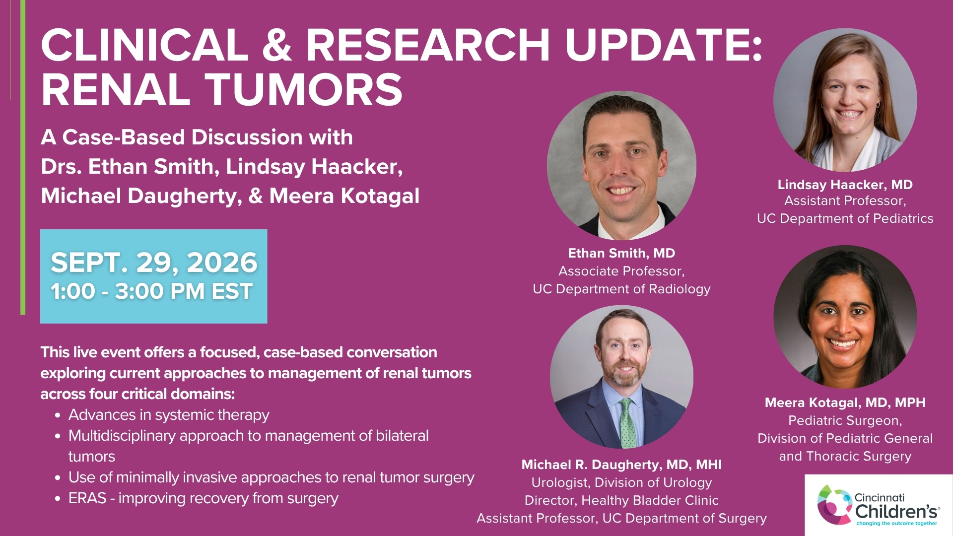
Clinical & Research Update: Renal Tumors with Drs. Ethan Smith, Lindsay Haacker, Michael Daugherty, and Meera Kotagal
71 min · Published Sep 2026
Video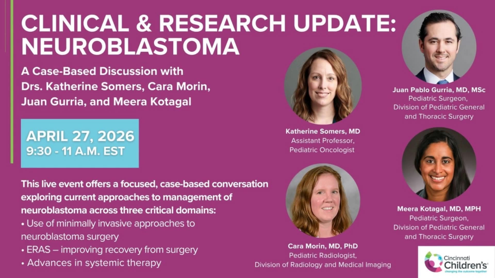
Clinical & Research Update: Neuroblastoma with Drs. Katherine Somers, Cara Morin, Juan Gurria, and Meera Kotagal
67 min · Published Sep 2026
Video
Clinical & Research Update: Sarcoma w/ Drs. Roshni Dasgupta, Joseph Pressey, Arthur Meyer, Luke Pater
66 min · Published Sep 2026
Video
2026 Laparoscopic Pediatric Hernia Repair
123 min · Published Jun 2026
Video
Beyond the Spectrum: Diagnosis, Myths & Management - Caitlin Couch & Leslie Lopez - APP Conference 2026
50 min · Published May 2026
Video
Dysautonomia: Navigating the Journey - Martha Willis - APP Conference 2026
55 min · Published May 2026
What the experts said
The complex category in the Children's Hospital study included any case with any other anomaly, including genetic anomalies (aneuploidies, trisomy 13/18), extracranial findings, and syndromic findings, creating a heterogeneous group.
Current counseling practice tends to combine isolated aqueductal stenosis cases (without suspected genetic cause) with complex cases in a broad-strokes approach, which may not be the appropriate approach.
Conventional endoscopes for ETV are usually about 6 millimeters in diameter.
The fetal ETV scope is a 2.4 mm sharp-tip fetoscope with a 1.3 mm camera and a working channel, the same equipment used for placing tracheal balloons in CDH fetuses.
The proposed gestational age for fetal ETV in phase one human studies is 24-25 weeks, following diagnosis around 20-21 weeks and genetic workup, similar to timing for spina bifida repair.
Intervention between 25 and 30 weeks gestational age is considered acceptable for cases with delayed diagnosis, allowing time for brain recovery in utero.
The fetal ETV trial requires an FDA IND (Investigational New Drug application) because it is a risky procedure, even though no drug is used and no device is left behind.
The goal is to begin a pilot study for safety and feasibility of fetal ETV next year, involving collaboration between fetal surgery and neurosurgery teams.
Acetazolamide can reduce CSF production by approximately 15% at best.
Acetazolamide may allow delaying intervention by one to two months in borderline, slowly progressing cases to reach a better category for ETV.
Acetazolamide has sufficient safety experience and electrolyte monitoring protocols to be used as a temporizing measure in appropriate patients.
Acetazolamide cannot be expected to be a game changer in rapidly progressing hydrocephalus cases.
Early postnatal ETV has a high failure rate in newborns with congenital aqueductal stenosis.
In a study by Rus, type 2 slowly progressive hydrocephalus patients treated with delayed ETV had the same neurologic outcomes as patients who were shunted earlier.
For stable postnatal patients not showing signs of rapid progression, there is no convincing data that earlier intervention is always better in a linear fashion.
For patients showing active progression with increasing pressure and damage, sooner intervention is better than waiting.
The primary benefit of fetal ETV may be developmental changes rather than preventing procedure failure, as many fetal ETV patients may still experience postnatal failure.
In postnatal treatment, the timing of intervention (earlier versus slightly later) is probably not as critical because the damage is already done and the condition is stable.
In fetal cases, timing is very critical because early intervention can potentially avoid pressure and allow better brain development.
The ETV fenestration can close in both neonates and fetuses, which is a concern for fetal intervention.
Maintaining decompression for 6-7 weeks in utero without pressure is beneficial even if postnatal redo ETV or shunt placement is needed, because the brain will have developed better.
The goal of fetal ETV is not to completely fix the hydrocephalus but to temporize and achieve better brain development, even if the intervention is temporary.
For fetal ETV cases, 2.4 millimeter scopes are being used in clinical studies.